Lab-Grown 3-D Brain Tissue Mimics Cortex

From cortical neurons, researchers have engineered rat tissue that formed complex networks of functioning neurons and appeared to behave normally after an injury.

Written byKerry Grens
| 1 min read

Register for free to listen to this article
Listen with Speechify
0:00
1:00
Share

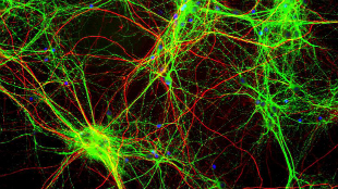
WIKIMEDIA, GERRYSHAWScientists have taken cortical neurons from a rat, seeded them onto a silk fiber-based scaffold, and assembled the structures into 3-D, donut-shape constructs. The end result is a chunk of tissue largely resembling functional brain matter, scientists reported in PNAS today (August 11).

“This work is an exceptional feat,” Rosemarie Hunziker, the program director of Tissue Engineering at the National Institute of Biomedical Imaging and Bioengineering, which helped fund the research, said in a press release. “It combines a deep understand [sic] of brain physiology with a large and growing suite of bioengineering tools to create an environment that is both necessary and sufficient to mimic brain function.”

David Kaplan of Tufts University led the group that developed the technology. The team built a ring-shape scaffold out of fibroin, a protein found in silk. Six concentric scaffold circles—each seeded with a different neuronal cell type—represented the six layers of the mammalian cortex. “It’s a form-fitting, Lego-like system, so we don’t have to worry about using glues, and how ...

Interested in reading more?

Become a Member of

The Scientist Logo
Receive full access to more than 35 years of archives, as well as TS Digest, digital editions of The Scientist, feature stories, and much more!
Already a member? Login Here

Related Topics

Meet the Author

  • kerry grens

    Kerry served as The Scientist’s news director until 2021. Before joining The Scientist in 2013, she was a stringer for Reuters Health, the senior health and science reporter at WHYY in Philadelphia, and the health and science reporter at New Hampshire Public Radio. Kerry got her start in journalism as a AAAS Mass Media fellow at KUNC in Colorado. She has a master’s in biological sciences from Stanford University and a biology degree from Loyola University Chicago.

    View Full Profile
Share
Image of a woman with her hands across her stomach. She has a look of discomfort on her face. There is a blown up image of her stomach next to her and it has colorful butterflies and gut bacteria all swarming within the gut.
November 2025, Issue 1

Why Do We Feel Butterflies in the Stomach?

These fluttering sensations are the brain’s reaction to certain emotions, which can be amplified or soothed by the gut’s own “bugs".

View this Issue
Olga Anczukow and Ryan Englander discuss how transcriptome splicing affects immune system function in lung cancer.

Long-Read RNA Sequencing Reveals a Regulatory Role for Splicing in Immunotherapy Responses

Pacific Biosciences logo
Research Roundtable: The Evolving World of Spatial Biology

Research Roundtable: The Evolving World of Spatial Biology

Conceptual cartoon image of gene editing technology

Exploring the State of the Art in Gene Editing Techniques

Bio-Rad
Conceptual image of a doctor holding a brain puzzle, representing Alzheimer's disease diagnosis.

Simplifying Early Alzheimer’s Disease Diagnosis with Blood Testing

fujirebio logo

Products

Labvantage Logo

LabVantage Solutions Awarded $22.3 Million U.S Customs and Border Protection Contract to Deliver Next-Generation Forensic LIMS

The Scientist Placeholder Image

Evosep Unveils Open Innovation Initiative to Expand Standardization in Proteomics

OGT logo

OGT expands MRD detection capabilities with new SureSeq Myeloid MRD Plus NGS Panel