
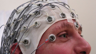
 Electrodes are held in place with a cap during an EEG recordingWIKIMEDIA, CHRIS HOPEGeneral anesthesia has transformed surgery from a ghastly ordeal to a procedure in which the patient feels no pain. Despite its widespread use, however, little is known about how anesthesia produces loss of consciousness—a blind spot brought into sharp focus by the fact that patients still occasionally wake up during surgery. But over the past 5 years, researchers have made significant progress in understanding what happens in the brain as consciousness departs and returns.
Peering into the anesthetized brain with neuroimaging and electroencephalograph (EEG) recordings, scientists have found evidence to support the “integrated-information theory,” which holds that consciousness relies on communication between different brain areas, and fades as that communication breaks down. EEG studies have also revealed distinctive brain wave patterns that signal when consciousness is lost and regained, offering easily identifiable markers for this impairment of communication.
Though many questions remain, advances in brain activity monitoring promise to shed light the neural basis of consciousness, and to eradicate the nightmare of mid-surgery awakenings. What’s more, by combining EEGs with magnetic brain stimulation, researchers may be able to measure consciousness and track recovery in unresponsive patients diagnosed as “vegetative,” who in recent years have been shown to sometimes have ...


