Molecular Motors

Researchers control nanomotors inside living cells.

Written byAbby Olena, PhD
| 1 min read

Register for free to listen to this article
Listen with Speechify
0:00
1:00
Share

MALLOUK LAB, PENN STATE UNIVERSITYManipulating tiny machines inside the body for diagnosing or treating illness is a goal that medicine has not yet achieved, but new research takes scientists one step closer. A team from Penn State University has safely manipulated nanomotors in living cells, according to a study published online today (February 12) in Angewandte Chemie.

“Our first-generation motors required toxic fuels and they would not move in biological fluid, so we couldn’t study them in human cells,” project leader Thomas Mallouk said in a statement. “That limitation was a serious problem.”

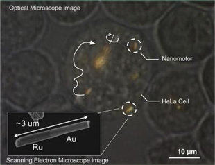
Mallouk and his colleagues addressed this issue by creating the nanomotors out of gold rods—approximately 300 nanometers in diameter and three microns long—and powering them using ultrasound, which at low power is non-toxic. They incubated the nanomotors with HeLa cells, which internalized the motors after about 24 hours. The researchers used low-power ultrasound to make the nanomotors move and spin and controlled their directionality using magnets. After, they showed that the cells were still viable. When the researchers used ultrasound at higher power, they could selectively destroy the cells ...

Interested in reading more?

Become a Member of

The Scientist Logo
Receive full access to more than 35 years of archives, as well as TS Digest, digital editions of The Scientist, feature stories, and much more!
Already a member? Login Here

Related Topics

Meet the Author

  • abby olena

    As a freelancer for The Scientist, Abby reports on new developments in life science for the website. She has a PhD from Vanderbilt University and got her start in science journalism as the Chicago Tribune’s AAAS Mass Media Fellow in 2013. Following a stint as an intern for The Scientist, Abby was a postdoc in science communication at Duke University, where she developed and taught courses to help scientists share their research. In addition to her work as a science journalist, she leads science writing and communication workshops and co-produces a conversational podcast. She is based in Alabama.  

    View Full Profile
Share
Illustration of a developing fetus surrounded by a clear fluid with a subtle yellow tinge, representing amniotic fluid.
January 2026, Issue 1

What Is the Amniotic Fluid Composed of?

The liquid world of fetal development provides a rich source of nutrition and protection tailored to meet the needs of the growing fetus.

View this Issue
Skip the Wait for Protein Stability Data with Aunty

Skip the Wait for Protein Stability Data with Aunty

Unchained Labs
Graphic of three DNA helices in various colors

An Automated DNA-to-Data Framework for Production-Scale Sequencing

illumina
Exploring Cellular Organization with Spatial Proteomics

Exploring Cellular Organization with Spatial Proteomics

Abstract illustration of spheres with multiple layers, representing endoderm, ectoderm, and mesoderm derived organoids

Organoid Origins and How to Grow Them

Thermo Fisher Logo

Products

Brandtech Logo

BRANDTECH Scientific Introduces the Transferpette® pro Micropipette: A New Twist on Comfort and Control

Biotium Logo

Biotium Launches GlycoLiner™ Cell Surface Glycoprotein Labeling Kits for Rapid and Selective Cell Surface Imaging

Colorful abstract spiral dot pattern on a black background

Thermo Scientific X and S Series General Purpose Centrifuges

Thermo Fisher Logo
Abstract background with red and blue laser lights

VANTAstar Flexible microplate reader with simplified workflows

BMG LABTECH