No Mo’ Slow Flow

Tools and tricks for high-throughput flow cytometry

Written byJeffrey M. Perkel
| 7 min read

Register for free to listen to this article
Listen with Speechify
0:00
7:00
Share

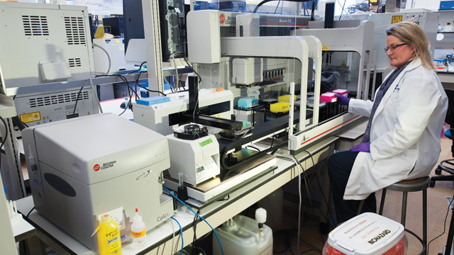
The assay setup used by the J. Paul Robinson lab at Purdue University Cytometry Laboratories to screen compounds for mitochondrial toxicity. PURDUE UNIVERSITY, MARK SIMONS

In the world of high-throughput screening, it’s all about efficiency: how many compounds can you test, at what cost, and using how little in the way of materials? In such an environment, cell-free and cell-based microtiter plate assays, such as ELISA and cell imaging, typically hold sway, as these are relatively cheap, low-volume, and amenable to automation.

One technology that has not usually been associated with high-throughput screening is flow cytometry, which traditionally has been long on content—the amount of data points possible per cell—but short on throughput—the number of samples that can be run in a day. But that, says J. Paul Robinson, director of Purdue University Cytometry Laboratories, is changing. As a screening platform, Robinson says, flow cytometry “is starting to gain some ...

Interested in reading more?

Become a Member of

The Scientist Logo
Receive full access to digital editions of The Scientist, as well as TS Digest, feature stories, more than 35 years of archives, and much more!
Already a member? Login Here

Related Topics

Meet the Author

Published In

Share
Illustration of a developing fetus surrounded by a clear fluid with a subtle yellow tinge, representing amniotic fluid.
January 2026, Issue 1

What Is the Amniotic Fluid Composed of?

The liquid world of fetal development provides a rich source of nutrition and protection tailored to meet the needs of the growing fetus.

View this Issue
Skip the Wait for Protein Stability Data with Aunty

Skip the Wait for Protein Stability Data with Aunty

Unchained Labs
Graphic of three DNA helices in various colors

An Automated DNA-to-Data Framework for Production-Scale Sequencing

illumina
Exploring Cellular Organization with Spatial Proteomics

Exploring Cellular Organization with Spatial Proteomics

Abstract illustration of spheres with multiple layers, representing endoderm, ectoderm, and mesoderm derived organoids

Organoid Origins and How to Grow Them

Thermo Fisher Logo

Products

Brandtech Logo

BRANDTECH Scientific Introduces the Transferpette® pro Micropipette: A New Twist on Comfort and Control

Biotium Logo

Biotium Launches GlycoLiner™ Cell Surface Glycoprotein Labeling Kits for Rapid and Selective Cell Surface Imaging

Colorful abstract spiral dot pattern on a black background

Thermo Scientific X and S Series General Purpose Centrifuges

Thermo Fisher Logo
Abstract background with red and blue laser lights

VANTAstar Flexible microplate reader with simplified workflows

BMG LABTECH