

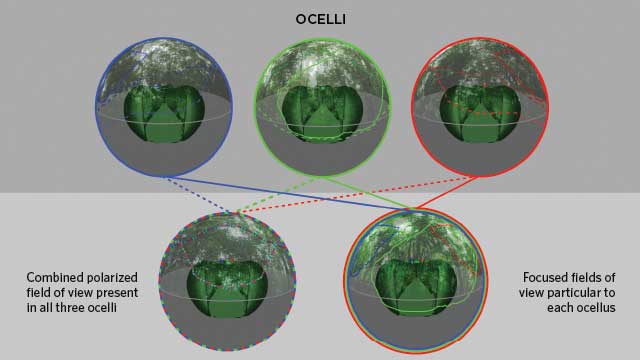
 THREE IN ONE: The three ocelli (top row), or simple eyes, of an orchid bee contain two fields of view. The polarized-light field of view is shared among all three ocelli to provide a trinocular view of the sky (dashed lines in bottom row, left), which potentially acts as a navigational aid as the bee flies through its dense, canopy-covered rainforest habitat. The solid lines represent each ocellus’s second, unique field of view produced by the perception of focused light.CURR BIOL, doi:10.1016/j.cub.2016.03.038, 2016. REPRODUCED WITH PERMISSION.
The paper
G.J. Taylor et al., “The dual function of orchid bee ocelli as revealed by X-ray microtomography,” Curr Biol, doi:10.1016/j.cub.2016.03.038, 2016.
For neuroscientist Emily Baird, a researcher who describes her passions as “animals, behavior, flying, and robots,” bees have always offered a fascinating study system. But in recent years, Baird, the head of a research team at Lund University in Sweden, has become particularly interested in how flight mechanisms are molded by the environments in which they operate.
While honeybees and bumblebees fly through relatively open habitats, for instance, decelerating to land on appealing flowers, other species, such as orchid bees (Euglossa imperialis), hurtle through the cluttered and ill-lit undergrowth of tropical rainforests. “I was interested in understanding how a bee that lives in ...


