
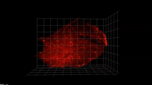
 3-D visualization of thymus vasculature in mice on day 14 after a non-lethal dose of radiation. The volume of vasculature in the thymus steadily decreased after damage.T. WERTHEIMER ET AL., SCIENCE IMMUNOLOGY, DOI:10.1126/sciimmunol.aal2736, 2017.The thymus, an organ in the lymphatic system, plays a critical role in immune function, producing the T cells essential to the immune response. While it is susceptible to damage, it also has the ability to repair itself via mechanisms that are not entirely clear. In a study published today (January 12) in Science Immunology, researchers identified a growth factor protein, BMP4, which is produced by endothelial cells in the inner wall of the thymus and is critical for its repair in mice. They also found that injecting these cells either into the blood stream or into the thymus itself sped up thymus recovery.
The study’s authors identified a new axis in thymus regeneration, says Avinash Bhandoola, head of the T-Cell Biology and Development Unit at the National Cancer Institute’s Center for Cancer Research, who was not involved in the study. “They showed that endothelial cells make this molecule BMP4, and this is actually really important for accelerating regeneration of the thymus after damage.”
The thymus, which gets smaller as we age, is highly sensitive to damage from stress and infection. And while it can recover from such insults—the process is known as endogenous thymic regeneration—more serious injury, for example, from chemotherapy or radiation, can extend recovery time considerably. That can result in an increased susceptibility to infections and even cancer relapse in patients while their T-cell count is ...


