

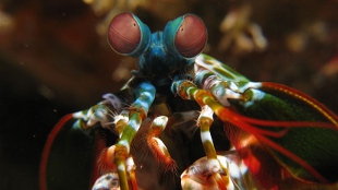
 Odontodactylus scyllarus, a mantis shrimp species closely related to the one studied by Bok et al.WIKIMEDIA, PRILFISHMantis shrimp (Neogonodactylus oerstedii) have 16 different types of photoreceptors in their eyes, and six of them are ultraviolet (UV) receptors. According to a study published online in Current Biology on July 3, these specialized receptors are each spectrally tuned to a different ultraviolet frequency by overlying filters composed of various mycosporine-like amino acid (MAA) pigments, which are typically found in the skin of marine organisms and function to block harmful UV rays.
Researchers had found previously that mantis shrimp do not have better color vision than other animals that can sense color (humans typically have three color photoreceptors), but researchers Lund University in Sweden and the University of South Dakota have discovered that the crustaceans have a remarkable ability to sense UV light. Though mantis shrimp contain only two types of visual pigments, called opsins, in their UV-sensitive receptors, they are able to sense many wavelengths of UV light by employing different UV blocking filters in conjunction with the photoreceptors. “The UV filters block certain wavelengths of light from reaching the photoreceptors, chromatically shifting their sensitivity,” lead author Michael Bok, a biologist at Lund University, said in a press release. “The effect is akin to putting red-tinted ...


