Microscopy image of a hair follicle shaft with clusters of orange debris surrounded by a group of green cells. Purple cells surround the hair root.
| 2 min read
In hair follicles, as nearby cells die, stem cells engage in phagocytosis to keep the hair cycle going.

Science Snapshot

A cross section of the pistil of <em >Arabidopsis thaliana&nbsp;</em>plant. Pollen grains are labeled with fluorescent markers and sit at the top of the structure. Fluorescently tagged pollen tubes penetrate the plant&rsquo;s ovary where the ovules (small, curved structures) are located.

The Hidden Dance of Plant Fertilization

Image of aggregated anthrobots (green) forming a bridge between two neuronal cells (red).

Building Living Bridges with Anthrobots

Neurons in culture

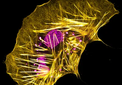
Lighting Up the Neuronal Cytoskeleton

Larger green cell with two cyan spots and many smaller magenta circles in and around the cell.&nbsp;

Synthetic Systems for Studying Natural Cells

An antenna-like cellular structure emerges from the surface of a human pancreatic islet cell. The surface of the cell is covered with small structures called microvilli.

The Shape of Cilia

Image of a brain section with various colors to denote different cell types (left) and different brain regions (right).

A Cell-by-Cell Map of the Entire Mouse Brain

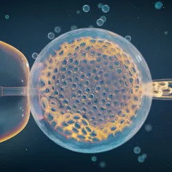
Fluorescent microscopy image of an embryo model representing four tissue layers of this developmental stage. Cyan-labeled cells are clustered at the top of an ovoid structure, underneath them are yellow-labeled cells and under them red-labeled cells, and surrounding all three layers are white labeled cells.

The First Human Embryo Model From Embryonic Stem Cells

The mouse peripheral nervous system with nerve cells farther away from the camera represented in yellow and pink, while nerves closer to the camera are shown in blue tones.

A Glowing Mouse Map

Image of pancreatic organoids under a microscope with immunofluorescent staining

Pancreatic Organoids Take the Stage

Fluorescently labeled neuron cell bodies in blue in the center compartment of a three-compartment microfluidic chamber grow through tiny grooves to enter the left and the right chambers, where they extend axons fibers, also shown in blue.

Visualizing Axon Pruning

This is DishBrain, an in vitro system that combines human brain cells with artificial intelligence technology, which researchers at Australian biotech Cortical Labs recently trained to play the classic 1972 Atari video game Pong. Fluorescent markers show axons in green, neuron bodies in purple, dendrites in red, and all other cells in blue. Where multiple markers are present, colors are merged and typically appear as yellow or pink. Posted: October 12, 2022

Caught on Camera

An international team of scientists used sensitive instruments to show that oil from human skin reacts with ozone to generate potent, free radicals. These chemicals can further react with most organic compounds present in the indoor environment to produce dangerous pollutants.&nbsp;

Caught on Camera

This young crustacean (Idotea balthica) carries the stained spermatia (blue) of the red algae Gracilaria gracilis on its body, marking the first known example of an animal facilitating fertilization in this ancient photosynthetic lineage.

Caught on Camera

Artist&rsquo;s rendition of <em>Meraxes gigas</em>&rsquo;s head

Caught on Camera

orange tiny frog

Caught on Camera

A female juvenile vaquitas, photographed in 2017.

Caught on Camera

Trichaptum abietinum growing on a pine log

Caught on Camera

zebrafish (Danio rerio)

Caught on Camera

A close-up of the eyespot on the wing of a forest mother-of-pearl butterfly (Protogoniomorpha parhassus)

Caught on Camera

Equid burial from Umm el-Marra, Syria

Caught on Camera

Page 1 of 97 - 1939 Total Items

Trending

Photo of John Calhoun crouches within his rodent utopia-turned-dystopia

Universe 25 Experiment

A profile of a young child with a measles rash.

Immune Amnesia: How the Texas Measles Outbreak Could Promote the Spread of Other Infectious Diseases

A red male cardinal and a brownish female sit on a branch in front of a green background.

Sex Determination: It’s in the Genes—Sometimes

An artistic rendition of polycystic ovarian syndrome, showing ovarian cysts represented by paper balls.

An Anti-Malarial Drug Could Ameliorate PCOS Symptoms

Multimedia

Olink Logo

Olink® Reveal: Accessible NGS-based proteomics for every lab

Olink logo
A researcher’s gloved hand holding a glass weighing boat with a loose powder sample on it

Minimizing to Maximize Measurement Accuracy

sartorius logo
A greyscale image of cells dividing.
March 2025, Issue 1

How Do Embryos Know How Fast to Develop

In mammals, intracellular clocks begin to tick within days of fertilization.

View this Issue
Discover the history, mechanics, and potential of PCR.

Become a PCR Pro

Integra Logo
3D rendered cross section of influenza viruses, showing surface proteins on the outside and single stranded RNA inside the virus

Genetic Insights Break Infectious Pathogen Barriers

Thermo Fisher Logo
A photo of sample storage boxes in an ultra-low temperature freezer.

Navigating Cold Storage Solutions

PHCbi logo&nbsp;
The Immunology of the Brain

The Immunology of the Brain

Products

Sapio Sciences

Sapio Sciences Makes AI-Native Drug Discovery Seamless with NVIDIA BioNeMo

DeNovix Logo

New DeNovix Helium Nano Volume Spectrophotometer

Olink Logo

Olink® Reveal: Accessible NGS-based proteomics for every lab

Olink logo
Zymo Logo

Zymo Research Launches the Quick-16S™ Full-Length Library Prep Kit