Viral Skeleton

A newly discovered family of tubulins—members of the cytoskeleton—encoded by bacteriophages plays a role in arranging the location of DNA within virus’s bacterial host.

Written byKerry Grens
| 2 min read

Register for free to listen to this article
Listen with Speechify
0:00
2:00
Share

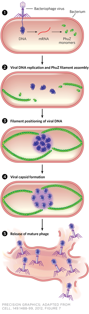
PhuZed Phage: When phage infect bacteria, host proteins begin transcribing a viral gene to produce PhuZ proteins (1), which form tubulin-like polymers that elongate from each pole of the cell (2). The filaments position replicating viral DNA at the cell’s center (3). Newly made viral DNA is encased in capsids (4), and released by lysis from the cell after final assembly (5) to infect other bacteria.

THE PAPERJ.A. Kraemer et al., “A phage tubulin assembles dynamic filaments by an atypical mechanism to center viral DNA within the host cell,” Cell, 149:1488-99, 2012.Until a few years ago, the cytoskeleton was thought to be a structure unique to eukaryotes, but a stream of discoveries in the past decade has shown that prokaryotes also have actin- and tubulin-like components. Tubulins are proteins that form microtubules, which manage everything from sorting genetic material during cell division to supporting cell shape and assisting in cell motility. A collaboration between David Agard’s group at the University of California, San Francisco, and Joe Pogliano’s lab at UC San Diego has now shown that a bacteriophage virus also encodes a family of tubulin-like proteins, which appear to determine the ...

Interested in reading more?

Become a Member of

The Scientist Logo
Receive full access to digital editions of The Scientist, as well as TS Digest, feature stories, more than 35 years of archives, and much more!
Already a member? Login Here

Related Topics

Meet the Author

  • kerry grens

    Kerry served as The Scientist’s news director until 2021. Before joining The Scientist in 2013, she was a stringer for Reuters Health, the senior health and science reporter at WHYY in Philadelphia, and the health and science reporter at New Hampshire Public Radio. Kerry got her start in journalism as a AAAS Mass Media fellow at KUNC in Colorado. She has a master’s in biological sciences from Stanford University and a biology degree from Loyola University Chicago.

    View Full Profile

Published In

Share
Illustration of a developing fetus surrounded by a clear fluid with a subtle yellow tinge, representing amniotic fluid.
January 2026, Issue 1

What Is the Amniotic Fluid Composed of?

The liquid world of fetal development provides a rich source of nutrition and protection tailored to meet the needs of the growing fetus.

View this Issue
Skip the Wait for Protein Stability Data with Aunty

Skip the Wait for Protein Stability Data with Aunty

Unchained Labs
Graphic of three DNA helices in various colors

An Automated DNA-to-Data Framework for Production-Scale Sequencing

illumina
Exploring Cellular Organization with Spatial Proteomics

Exploring Cellular Organization with Spatial Proteomics

Abstract illustration of spheres with multiple layers, representing endoderm, ectoderm, and mesoderm derived organoids

Organoid Origins and How to Grow Them

Thermo Fisher Logo

Products

Brandtech Logo

BRANDTECH Scientific Introduces the Transferpette® pro Micropipette: A New Twist on Comfort and Control

Biotium Logo

Biotium Launches GlycoLiner™ Cell Surface Glycoprotein Labeling Kits for Rapid and Selective Cell Surface Imaging

Colorful abstract spiral dot pattern on a black background

Thermo Scientific X and S Series General Purpose Centrifuges

Thermo Fisher Logo
Abstract background with red and blue laser lights

VANTAstar Flexible microplate reader with simplified workflows

BMG LABTECH