

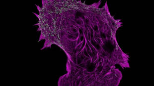
 Filamentous actin, captured by super-resolution structured illumination microscopy VIMEO, HHMI NEWSEric Betzig, Stefan Hell, and William Moerner scooped up the 2014 Nobel Prize in Chemistry for their contributions to the development of super-resolved fluorescence microscopy, which broke the theoretical limit of microscopic resolution imposed by the wavelength of light. Now, Betzig, of Janelia Research Campus in Ashburn, Virginia, and his colleagues have applied the new techniques to watching live cells in action, generating images and videos of protein movement and interactions as the cells internalize molecules. The team published its results this week (August 27) in Science.
“These methods set a new standard for how far you can push the speed and non-invasiveness of super-resolution imaging,” Betzig said in a press release. “This will bring super-resolution to live-cell imaging for real.”
Betzig and his colleagues achieved their success by improving the spatial resolution of structured illumination microscopy (SIM). With traditional SIM, images are generated by switching on the fluorescent labels that researchers have used to tag specific proteins, followed by a wave of light that deactivates most of them. The tags in the darkest regions continue to fluoresce, however, sharpening the image. Repeating this process more than two dozen times can yield a high-resolution composite image. But the time it takes to switch the tags on and off has made the technique ...


