Zika and Birth Defects

A study provides stronger evidence for the causal link between Zika infection and microcephaly, and warns of a host of other birth defects associated with the virus.

Written byCatherine Offord
| 2 min read

Register for free to listen to this article
Listen with Speechify
0:00
2:00
Share

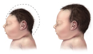
A baby with microcephaly (left) compared to a baby with a typical-sized head (right)WIKIMEDIA, CDCMore than one-quarter of women taking part in a study of Zika infection during pregnancy had fetuses with potentially serious abnormalities, according to a report published last week (March 4) in the New England Journal of Medicine. The analysis compared fetuses in infected mothers to fetuses in non-infected mothers, thereby providing the control-like group that many previous studies have lacked.

The analysis is “what people have been waiting for,” Amesh Adalja of the University of Pittsburgh Medical Center, who was not involved in the work, told LiveScience, adding that the comparison of the two groups allowed stronger links to be drawn between Zika infection and microcephaly. “This is the closest we’ve gotten to [proving] causation. . . . For all intents and purposes, this justifies the concern raised early on.”

Of the 88 women enrolled in the study, 72 tested positive for Zika virus infection. While examination of the fetuses of all Zika-negative women found no defects, examinations of the fetuses in 42 Zika-infected women revealed abnormalities in 12, including central nervous system lesions (seven fetuses) and growth restriction with or without microcephaly (five fetuses). ...

Interested in reading more?

Become a Member of

The Scientist Logo
Receive full access to more than 35 years of archives, as well as TS Digest, digital editions of The Scientist, feature stories, and much more!
Already a member? Login Here

Related Topics

Meet the Author

  • After undergraduate research with spiders at the University of Oxford and graduate research with ants at Princeton University, Catherine left arthropods and academia to become a science journalist. She has worked in various guises at The Scientist since 2016. As Senior Editor, she wrote articles for the online and print publications, and edited the magazine’s Notebook, Careers, and Bio Business sections. She reports on subjects ranging from cellular and molecular biology to research misconduct and science policy. Find more of her work at her website.

    View Full Profile
Share
Illustration of a developing fetus surrounded by a clear fluid with a subtle yellow tinge, representing amniotic fluid.
January 2026, Issue 1

What Is the Amniotic Fluid Composed of?

The liquid world of fetal development provides a rich source of nutrition and protection tailored to meet the needs of the growing fetus.

View this Issue
Skip the Wait for Protein Stability Data with Aunty

Skip the Wait for Protein Stability Data with Aunty

Unchained Labs
Graphic of three DNA helices in various colors

An Automated DNA-to-Data Framework for Production-Scale Sequencing

illumina
Exploring Cellular Organization with Spatial Proteomics

Exploring Cellular Organization with Spatial Proteomics

Abstract illustration of spheres with multiple layers, representing endoderm, ectoderm, and mesoderm derived organoids

Organoid Origins and How to Grow Them

Thermo Fisher Logo

Products

Brandtech Logo

BRANDTECH Scientific Introduces the Transferpette® pro Micropipette: A New Twist on Comfort and Control

Biotium Logo

Biotium Launches GlycoLiner™ Cell Surface Glycoprotein Labeling Kits for Rapid and Selective Cell Surface Imaging

Colorful abstract spiral dot pattern on a black background

Thermo Scientific X and S Series General Purpose Centrifuges

Thermo Fisher Logo
Abstract background with red and blue laser lights

VANTAstar Flexible microplate reader with simplified workflows

BMG LABTECH