Zooming In

To improve the reach of optical microscopy, researchers are enlarging the biological features they wish to view.

Written byJef Akst
| 1 min read

Register for free to listen to this article
Listen with Speechify
0:00
1:00
Share

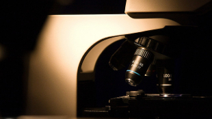
FLICKR, MILOSZ1By enlarging cells and tissues by up five times their normal size, MIT researchers were able to observe some of the smallest features of life—such as individual neurons and synapses—using traditional optical microscopes, according to a study published last week (January 15) in Science. Edward Boyden, codirector of the MIT Center for Neurobiological Engineering, and his colleagues used the new technique, called expansion microscopy, to view objects as small as 70 nanometers—well below the typical 200-nanometer limit of conventional optical microscopes.

“We hope we have a technology that will allow you to scan the nervous system of entire animals,” Boyden told The New York Times (NYT).

The technique hinges on a polymer that is commonly found in diapers. Absorbing up to 300 times its mass in water, the material has the potential to cause biological entities to swell. By fluorescently tagging the structures of interest, then infusing the tissue with the composite parts of the polymer and adding water, the researchers were able to force the tissue to expand uniformly in all directions—up to five times its original size—while maintaining its overall organization. Boyden hopes that more fine-tuning could eventually result in the ability to expand tissues by 10 times or more, he told NYT.

Interested in reading more?

Become a Member of

The Scientist Logo
Receive full access to more than 35 years of archives, as well as TS Digest, digital editions of The Scientist, feature stories, and much more!
Already a member? Login Here

Related Topics

Meet the Author

  • Jef (an unusual nickname for Jennifer) got her master’s degree from Indiana University in April 2009 studying the mating behavior of seahorses. After four years of diving off the Gulf Coast of Tampa and performing behavioral experiments at the Tennessee Aquarium in Chattanooga, she left research to pursue a career in science writing. As The Scientist's managing editor, Jef edited features and oversaw the production of the TS Digest and quarterly print magazine. In 2022, her feature on uterus transplantation earned first place in the trade category of the Awards for Excellence in Health Care Journalism. She is a member of the National Association of Science Writers.

    View Full Profile
Share
Illustration of a developing fetus surrounded by a clear fluid with a subtle yellow tinge, representing amniotic fluid.
January 2026

What Is the Amniotic Fluid Composed of?

The liquid world of fetal development provides a rich source of nutrition and protection tailored to meet the needs of the growing fetus.

View this Issue
Human-Relevant In Vitro Models Enable Predictive Drug Discovery

Advancing Drug Discovery with Complex Human In Vitro Models

Stemcell Technologies
Redefining Immunology Through Advanced Technologies

Redefining Immunology Through Advanced Technologies

Ensuring Regulatory Compliance in AAV Manufacturing with Analytical Ultracentrifugation

Ensuring Regulatory Compliance in AAV Manufacturing with Analytical Ultracentrifugation

Beckman Coulter Logo
Conceptual multicolored vector image of cancer research, depicting various biomedical approaches to cancer therapy

Maximizing Cancer Research Model Systems

bioxcell

Products

Refeyn logo

Refeyn named in the Sunday Times 100 Tech list of the UK’s fastest-growing technology companies

Parse Logo

Parse Biosciences and Graph Therapeutics Partner to Build Large Functional Immune Perturbation Atlas

Sino Biological Logo

Sino Biological's Launch of SwiftFluo® TR-FRET Kits Pioneers a New Era in High-Throughout Kinase Inhibitor Screening

SPT Labtech Logo

SPT Labtech enables automated Twist Bioscience NGS library preparation workflows on SPT's firefly platform