ADVERTISEMENT
A pile of superworms.
The Culprit of a Mysterious Superworm Epidemic Finally Identified
Advanced microscopy aided in the detection of the pathogen responsible for a nationwide superworm apocalypse and informed the development of a potential vaccine strategy.
The Culprit of a Mysterious Superworm Epidemic Finally Identified

Advanced microscopy aided in the detection of the pathogen responsible for a nationwide superworm apocalypse and informed the development of a potential vaccine strategy.

Advanced microscopy aided in the detection of the pathogen responsible for a nationwide superworm apocalypse and informed the development of a potential vaccine strategy.

microscopy

Explore How Stains and Probes Work to Label Cellular Morphology
Cellular Stains Under the Hood
Biotium | Oct 1, 2024 | 1 min read
Fluorescent probes are key tools for scientists looking at cellular properties or morphology. Use this guide to determine which probe is best for a given situation or experiment.
A composite image showing raw capture of endosomes using lattice light sheet microscopy, represented by teal and purple spheres, and detection and tracking of endosomal movement, represented by teal and purple lines. 
Changing Identities: Imaging Endosomal Maturation
Nathan Ni, PhD | Aug 1, 2024 | 3 min read
Harrison York combines microscopy and machine learning to visualize and characterize how endosomes regulate intracellular mechanisms. 
A microscope and a model of a molecule, showcasing the power of atomic force microscopy for surface imaging at atomic resolution.
Atomic Force Microscopy to See the Invisible
Priyom Bose, PhD | 8 min read
Atomic force microscopy (AFM) is a technique for visualizing a variety of samples, from single molecules to living cells, at ultrahigh resolution. 
Neurons in culture
Lighting Up the Neuronal Cytoskeleton
Mariella Bodemeier Loayza Careaga, PhD | Aug 1, 2024 | 2 min read
By combining microscopy techniques with genome engineering, scientists revealed the complexities of the presynaptic actin cytoskeleton.
3D illustration of purple neurons.
An Immune Mechanism Maintains Memory
Shelby Bradford, PhD | Jun 10, 2024 | 4 min read
A receptor that recognizes DNA fragments formed during learning is necessary for proper memory formation.
Microscopic image of a live amoeba.
Illuminating Specimens Through Live Cell Imaging
Charlene Lancaster, PhD | 8 min read
Live cell imaging is a powerful microscopy technique employed by scientists to monitor molecular processes and cellular behavior in real time.
A 3D microscopic image of a heart tissue section with cardiac myocytes and macrophages.
Taking Out the Trash: An Alternative Cellular Disposal Pathway
Charlene Lancaster, PhD | Jun 6, 2024 | 4 min read
Researchers reveal that cellular secretion removes defective mitochondria when lysosomes are dysfunctional.
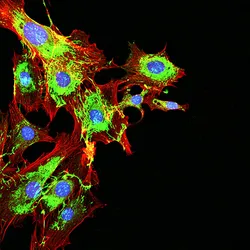
Image of fibroblast cell with nuclei (yellow), mitochondria (red)<br >, and microfilaments (blue).
Complicated CAP Does It All
Rachael Moeller Gorman | Apr 5, 2024 | 5 min read
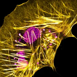
Researchers found that actin filaments can behave counter to decades-old actin dynamics dogma, changing how biologists think about cell movement.
Beyond Skin Deep: Analyzing Melanoma Cells Through Morphology
The Scientist | 1 min read
In this webinar, Evelyn Lattmann will discuss how she examined melanoma cell morphology without labels using an AI-based microscope system.  
The illustration shows gears inside a light bulb.
A Picture Sparks a Thousand Words
Meenakshi Prabhune, PhD | Oct 2, 2023 | 2 min read
A scientific image can conceal even more than it reveals. Scientists can now share their untold behind-the-image stories in our new Science Snapshot column.
Illustration showing origami tardigrade, fungi and bacteria.
Magnifying Curiosity with a Pocket Microscope
Danielle Gerhard, PhD | Sep 8, 2023 | 5 min read
Microscopes were inaccessible to most of the world until Manu Prakash and Jim Cybulski put their engineering prowess to the test.
<em >Immunohistochemistry: Origins, Tips, and a Look to the Future</em>
Immunohistochemistry: Origins, Tips, and a Look to the Future
Steven Hrycaj, PhD | 7 min read
An essential staining technique with a long history, immunohistochemistry (IHC) is being upgraded for modern research and clinical applications.
Infographic showing the difference between the classic MINFLUX and the updated MINFLUX
A New Kind of MINFLUX
Ida Emilie Steinmark, PhD | Sep 1, 2023 | 1 min read
MINFLUX brought record-breaking resolution to fluorescence microscopy in 2016. A new version perfected for protein tracking came out this spring.
Infographic showing a new way to assess antibiotic effectiveness based on how much bacteria jiggle
Jiggling Bacteria Reveal Antibiotic Resistance
Ida Emilie Steinmark, PhD | Jul 5, 2023 | 1 min read
Finding an effective antibiotic against an infection can easily take 24 hours. Faster testing could save lives and help doctors avoid broad-spectrum antibiotics, which can foster resistance.
Discover How CF&reg; Dyes Let Scientists Delve Deeper into Biological Phenomena
CF® Dyes: Clearer Fluorescent Results
Biotium | 1 min read
CF®  Dyes combine pegylation and sulfonation to create fluorescent dyes with better signal intensity, more solubility, and superior specificity.
Illustration of neuron cells network
A Complete Brain Wiring Map
Danielle Gerhard, PhD | Jul 5, 2023 | 2 min read
Scientists developed new tools for brain reconstruction and analysis to create and characterize a complete brain wiring map of the fruit fly larva for the first time.
Illustration of newly discovered mechanism allowing kinesin to &ldquo;walk&rdquo; down a microtubule. A green kinesin molecule with an attached yellow fluorophore is shown passing through a blue laser as it rotates step by step along a red and purple microtubule, fueled by blue ATP molecules that are hydrolyzed into orange ADP and phosphate groups.
High-Resolution Microscope Watches Proteins Strut Their Stuff
Holly Barker, PhD | Mar 31, 2023 | 3 min read
Modification on a high-resolution fluorescent microscopy technique allow researchers to track the precise movements of motor proteins. 
A blood sample containing white and red blood cells.
Enhancing Cell Morphology-Based Analysis
The Scientist and Deepcell | 3 min read
Learn how the latest AI-driven technology uses morphology to comprehensively analyze and sort cell populations.
A normal human liver organoid (left) stained with blue and red next to a fatty liver organoid (right) with lipid droplets stained yellow.
Working Together to Battle Fatty Liver Disorders
Niki Spahich, PhD | Mar 24, 2023 | 6 min read
Benedetta Artegiani and Delilah Hendriks formed a joint laboratory group to understand disease mechanisms and treatments through organoid models.
ADVERTISEMENT