Best Science Multimedia of 2011

A look at this year’s most memorable science videos, images, and websites

Written byCristina Luiggi
| 2 min read

Register for free to listen to this article
Listen with Speechify
0:00
2:00
Share

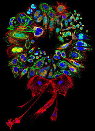
Dr. Donna Stolz, University of Pittsburgh Mammalian cell collage stained for various proteins and organelles, assembled into a wreath COURTESY OF NIKON SMALL WORLD

Nikon’s 2011 Small World CompetitionFor the past 37 years, Nikon’s Small World Photomicrography Competition has showcased some of the most breathtaking images ever captured under a microscope, and this year was no exception. The top three winners of this year’s competition were photographs of a turquoise-hued green lacewing, a blade of grass magnified 200 times, and the marine phytoplankton Melosira moniliformis, which was imaged while still alive.

Wellcome Image awards 2011Each year, Wellcome Images—a freely available online repository of historical and contemporary images from biology and medicine—recognize the most memorable images acquired into the collection that year. This year’s picks included a confocal micrograph of a mouse retina, a confocal micrograph of wheat infected with ergot fungus, and a stunning scanning electron micrograph of a ...

Interested in reading more?

Become a Member of

The Scientist Logo
Receive full access to more than 35 years of archives, as well as TS Digest, digital editions of The Scientist, feature stories, and much more!
Already a member? Login Here

Meet the Author

Share
Illustration of a developing fetus surrounded by a clear fluid with a subtle yellow tinge, representing amniotic fluid.
January 2026, Issue 1

What Is the Amniotic Fluid Composed of?

The liquid world of fetal development provides a rich source of nutrition and protection tailored to meet the needs of the growing fetus.

View this Issue
Skip the Wait for Protein Stability Data with Aunty

Skip the Wait for Protein Stability Data with Aunty

Unchained Labs
Graphic of three DNA helices in various colors

An Automated DNA-to-Data Framework for Production-Scale Sequencing

illumina
Exploring Cellular Organization with Spatial Proteomics

Exploring Cellular Organization with Spatial Proteomics

Abstract illustration of spheres with multiple layers, representing endoderm, ectoderm, and mesoderm derived organoids

Organoid Origins and How to Grow Them

Thermo Fisher Logo

Products

Brandtech Logo

BRANDTECH Scientific Introduces the Transferpette® pro Micropipette: A New Twist on Comfort and Control

Biotium Logo

Biotium Launches GlycoLiner™ Cell Surface Glycoprotein Labeling Kits for Rapid and Selective Cell Surface Imaging

Colorful abstract spiral dot pattern on a black background

Thermo Scientific X and S Series General Purpose Centrifuges

Thermo Fisher Logo
Abstract background with red and blue laser lights

VANTAstar Flexible microplate reader with simplified workflows

BMG LABTECH