Brighter, Smaller, Faster

As X-ray crystallography enters its second century, shrinking crystals and brighter light sources are redefining structural biology.

Written byJeffrey M. Perkel
| 7 min read

Register for free to listen to this article
Listen with Speechify
0:00
7:00
Share

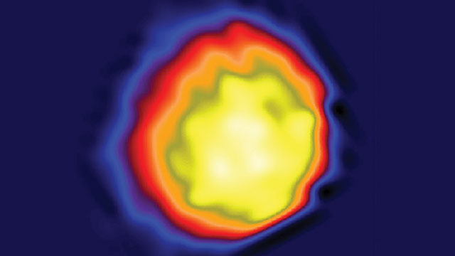
NAKED VIRUS: The first high-contrast image of a single herpesvirus ever taken with a coherent X-ray synchrotron. The virus, with its capsid, measures about 100 nm in diameter.COURTESY OF JIANWEI MIAO

It was just over a century ago that William Lawrence Bragg and his father, William Henry Bragg, kick-started the science of X-ray crystallography in a talk at the Cambridge Philosophical Society on November 11, 1912. Since then, thousands upon thousands of structures have been solved, from table salt and diamond to RNA polymerase and the ribosome.

Back in the Braggs’ time, crystals were analyzed using laboratory X-ray tubes, which are relatively weak sources of continuous, noncoherent light—that is, light that travels in all directions, like lamplight. In the 1970s, researchers started using synchrotron particle accelerators, which can shoot partially focused (i.e., relatively coherent), highly intense X-ray radiation at a crystal. According to Jianwei Miao, a professor of physics and astronomy at the University of California, ...

Interested in reading more?

Become a Member of

The Scientist Logo
Receive full access to digital editions of The Scientist, as well as TS Digest, feature stories, more than 35 years of archives, and much more!
Already a member? Login Here
Illustration of a developing fetus surrounded by a clear fluid with a subtle yellow tinge, representing amniotic fluid.
January 2026, Issue 1

What Is the Amniotic Fluid Composed of?

The liquid world of fetal development provides a rich source of nutrition and protection tailored to meet the needs of the growing fetus.

View this Issue
Skip the Wait for Protein Stability Data with Aunty

Skip the Wait for Protein Stability Data with Aunty

Unchained Labs
Graphic of three DNA helices in various colors

An Automated DNA-to-Data Framework for Production-Scale Sequencing

illumina
Exploring Cellular Organization with Spatial Proteomics

Exploring Cellular Organization with Spatial Proteomics

Abstract illustration of spheres with multiple layers, representing endoderm, ectoderm, and mesoderm derived organoids

Organoid Origins and How to Grow Them

Thermo Fisher Logo

Products

Brandtech Logo

BRANDTECH Scientific Introduces the Transferpette® pro Micropipette: A New Twist on Comfort and Control

Biotium Logo

Biotium Launches GlycoLiner™ Cell Surface Glycoprotein Labeling Kits for Rapid and Selective Cell Surface Imaging

Colorful abstract spiral dot pattern on a black background

Thermo Scientific X and S Series General Purpose Centrifuges

Thermo Fisher Logo
Abstract background with red and blue laser lights

VANTAstar Flexible microplate reader with simplified workflows

BMG LABTECH