Electrode-Free Electrophysiology

Optogenetics has evolved beyond its neuron-stimulating capacities to an all-optical approach for both manipulating and recording cells.

Written byKerry Grens
| 2 min read

Register for free to listen to this article
Listen with Speechify
0:00
2:00
Share

WIKIMEDIA, CHEMPETITIVETen years ago, optogenetics started out as a means to stimulate neuronal activity without the use of electrodes: a genetically introduced light-sensitive membrane channel and a blue light could do the job. At the Society for Neuroscience (SfN) meeting in Chicago this week, scientists announced new techniques for recording neurons optically, as well, obviating the need for invading cells or tissues with electrical probes altogether.

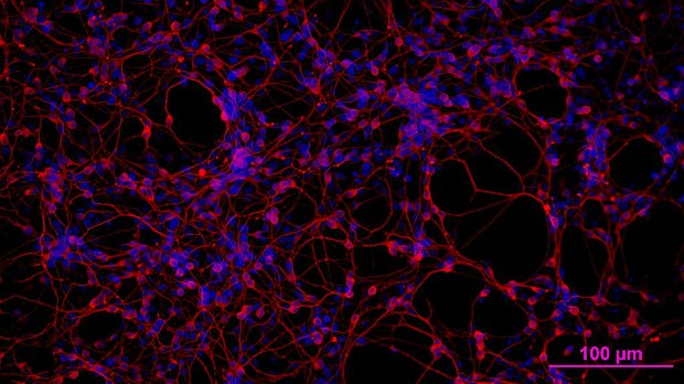
The basic approach is to express genes for a light-sensitive channel and an optical reporter in the same cell. For instance, researchers have succeeded in using genetically introduced calcium indicators, which signal action potentials, to record cell activity upon optogenetic stimulation.

To get an even more refined look at cell activity, particularly at the sub-action potential level, Adam Cohen’s team at Harvard University has developed an optical voltage indicator, called QuasAr, which glows in the near infrared upon changes in cellular voltage (voltage indicators act on a much faster time scale than calcium indicators). Along with Ed Boyden’s lab at MIT, Cohen developed a new channelrhodopsin that’s extremely sensitive to blue light and introduced it into cells along with QuasAr, allowing the researchers to stimulate ...

Interested in reading more?

Become a Member of

The Scientist Logo
Receive full access to more than 35 years of archives, as well as TS Digest, digital editions of The Scientist, feature stories, and much more!
Already a member? Login Here

Related Topics

Meet the Author

  • kerry grens

    Kerry served as The Scientist’s news director until 2021. Before joining The Scientist in 2013, she was a stringer for Reuters Health, the senior health and science reporter at WHYY in Philadelphia, and the health and science reporter at New Hampshire Public Radio. Kerry got her start in journalism as a AAAS Mass Media fellow at KUNC in Colorado. She has a master’s in biological sciences from Stanford University and a biology degree from Loyola University Chicago.

    View Full Profile
Share
Image of a woman with her hands across her stomach. She has a look of discomfort on her face. There is a blown up image of her stomach next to her and it has colorful butterflies and gut bacteria all swarming within the gut.
November 2025, Issue 1

Why Do We Feel Butterflies in the Stomach?

These fluttering sensations are the brain’s reaction to certain emotions, which can be amplified or soothed by the gut’s own “bugs".

View this Issue
Olga Anczukow and Ryan Englander discuss how transcriptome splicing affects immune system function in lung cancer.

Long-Read RNA Sequencing Reveals a Regulatory Role for Splicing in Immunotherapy Responses

Pacific Biosciences logo
Research Roundtable: The Evolving World of Spatial Biology

Research Roundtable: The Evolving World of Spatial Biology

Conceptual cartoon image of gene editing technology

Exploring the State of the Art in Gene Editing Techniques

Bio-Rad
Conceptual image of a doctor holding a brain puzzle, representing Alzheimer's disease diagnosis.

Simplifying Early Alzheimer’s Disease Diagnosis with Blood Testing

fujirebio logo

Products

Eppendorf Logo

Research on rewiring neural circuit in fruit flies wins 2025 Eppendorf & Science Prize

Evident Logo

EVIDENT's New FLUOVIEW FV5000 Redefines the Boundaries of Confocal and Multiphoton Imaging

Evident Logo

EVIDENT Launches Sixth Annual Image of the Year Contest

10x Genomics Logo

10x Genomics Launches the Next Generation of Chromium Flex to Empower Scientists to Massively Scale Single Cell Research