Embryonic Evolution Through Ernst Haeckel’s Eyes

The 19th century biologist’s drawings, tainted by scandal, helped bolster, then later dismiss, his biogenetic law.

Written byDiana Kwon
| 3 min read

Register for free to listen to this article
Listen with Speechify
0:00
3:00

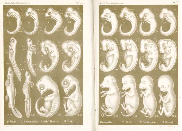
EMBRYONIC EVOLUTION: This comparative illustration of eight species’ embryos from Haeckel’s Anthropogenie (1874 edition) is among the most well-known of the German scientist’s images. The rows represent three developmental stages and the columns correspond to different species (fish, salamander, turtle, chicken, pig, cow, dog, and human). NICK HOPWOOD

Ernst Haeckel, a biologist, artist, and philosopher born in Prussia in the 1830s, played a key role in spreading Darwinism in Germany. He was also deeply fascinated by embryology and illustrated some of the most remarkable comparisons of vertebrate embryos in his day. These images were widely printed and copied, both to argue for Haeckel’s controversial evolutionary theories and to debunk them.

Haeckel’s most influential idea was his now-infamous biogenetic law, summarized by the phrase “ontogeny recapitulates phylogeny”—in other words, an organism’s embryo progresses through stages of development that mirror its evolutionary history. According to this theory, embryos of more advanced species—humans, for example—would pass through stages in which they displayed the adult characteristics of their more primitive ancestors (such as fish gills or monkey ...

Interested in reading more?

Become a Member of

The Scientist Logo
Receive full access to digital editions of The Scientist, as well as TS Digest, feature stories, more than 35 years of archives, and much more!
Already a member?
Add The Scientist as a preferred source on Google

Add The Scientist as a preferred Google source to see more of our trusted coverage.

Meet the Author

  • Diana is a freelance science journalist who covers the life sciences, health, and academic life. She’s a regular contributor to The Scientist and her work has appeared in several other publications, including Scientific American, Knowable, and Quanta. Diana was a former intern at The Scientist and she holds a master’s degree in neuroscience from McGill University. She’s currently based in Berlin, Germany.

    View Full Profile

Related Topics

Published In

May 2017

Rapid Evolution

Genetic change within populations can happen in mere generations

Related articles background image
The Scientist Digest cover September 2026
September 2026

Multiplex Microscopy Becomes Easier with Encoded Antibodies

A new system that enables researchers to uniquely tag monoclonal antibodies for use in microscopy could help simplify complex imaging studies.

View this Issue
Rethinking ALS Biomarkers: From Discovery to Clinical Impact

Rethinking ALS Biomarkers: From Discovery to Clinical Impact

Alamar Biosciences logo
Best Practices for qPCR Assay Design and Optimization

Best Practices for qPCR Assay Design and Optimization

Bio-Rad
Beyond the Basics: Strategies for Single-Cell and Spatial Transcriptomics Analysis

Beyond the Basics: Strategies for Single-Cell and Spatial Transcriptomics Analysis

bioxcell
Scientist reviewing cellular and molecular data on a computer in a laboratory.

Building Translation-Ready Biomarkers with Connected Workflows

Danaher Logo

Products

Closeup image of a multi channel pipette dispensing pink liquid into a 96-well plate.

The ASSIST PLUS pipetting robot for affordable workflow automation

Integra Logo
Single cells in suspension

Rapidly isolate primary cells and make uniform single-cell suspensions with Corning® Cell Strainers

Corning logo
Abstract image representing cell membranes linked together.

CellBrite® Steady Membrane Stain: Cell surface staining built for real-time imaging

Biotium
sino biological logo

Monod Bio Licenses AI-designed Protein Technologies to SignalChem Biotech for Custom Discovery Assays