Extracting Exosomes to Detect Signs of Cancer in Urine

A new device uses anchored nanowires to capture extracellular vesicles from pee for microRNA analysis.

Written byRuth Williams
| 2 min read

Register for free to listen to this article
Listen with Speechify
0:00
2:00
Share

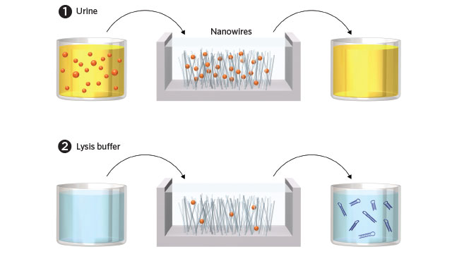
PEE IN, MICRORNAs OUT: (1) A small volume of urine is introduced into the microfluidic device, where positively charged zinc oxide nanowires attract and bind negatively charged exosomes. (2) Lysis buffer is introduced to the device to break open the exosomes and free the microRNAs, which are then collected for sequence analysis.

See full infographic: WEB© GEORGE RETSECK

Exosomes are tiny membrane-bound packages that are released from practically every cell type and found in a wide range of body fluids. Containing RNAs, proteins, and other cell components, they are believed to be involved in communication between cells, and there’s evidence that their abundance and content may change with disease state. Consequently, there is a growing interest in collecting and analyzing these vesicles for diagnostic purposes. Researchers who are interested in the diagnostic potential of microRNAs, for example, are especially keen to collect exosomes because the RNAs they contain degrade more slowly than free-floating RNAs.

“They are packed with ...

Interested in reading more?

Become a Member of

The Scientist Logo
Receive full access to digital editions of The Scientist, as well as TS Digest, feature stories, more than 35 years of archives, and much more!
Already a member? Login Here

Related Topics

Meet the Author

  • ruth williams

    Ruth is a freelance journalist. Before freelancing, Ruth was a news editor for the Journal of Cell Biology in New York and an assistant editor for Nature Reviews Neuroscience in London. Prior to that, she was a bona fide pipette-wielding, test tube–shaking, lab coat–shirking research scientist. She has a PhD in genetics from King’s College London, and was a postdoc in stem cell biology at Imperial College London. Today she lives and writes in Connecticut.

    View Full Profile

Published In

April 2018

Neuron Nets

Wrapped up in memory, addiction, and more

Share
Image of a woman with her hands across her stomach. She has a look of discomfort on her face. There is a blown up image of her stomach next to her and it has colorful butterflies and gut bacteria all swarming within the gut.
November 2025, Issue 1

Why Do We Feel Butterflies in the Stomach?

These fluttering sensations are the brain’s reaction to certain emotions, which can be amplified or soothed by the gut’s own “bugs".

View this Issue
Olga Anczukow and Ryan Englander discuss how transcriptome splicing affects immune system function in lung cancer.

Long-Read RNA Sequencing Reveals a Regulatory Role for Splicing in Immunotherapy Responses

Pacific Biosciences logo
Research Roundtable: The Evolving World of Spatial Biology

Research Roundtable: The Evolving World of Spatial Biology

Conceptual cartoon image of gene editing technology

Exploring the State of the Art in Gene Editing Techniques

Bio-Rad
Conceptual image of a doctor holding a brain puzzle, representing Alzheimer's disease diagnosis.

Simplifying Early Alzheimer’s Disease Diagnosis with Blood Testing

fujirebio logo

Products

Eppendorf Logo

Research on rewiring neural circuit in fruit flies wins 2025 Eppendorf & Science Prize

Evident Logo

EVIDENT's New FLUOVIEW FV5000 Redefines the Boundaries of Confocal and Multiphoton Imaging

Evident Logo

EVIDENT Launches Sixth Annual Image of the Year Contest

10x Genomics Logo

10x Genomics Launches the Next Generation of Chromium Flex to Empower Scientists to Massively Scale Single Cell Research