Flatworms’ Synapses Steer Body Shape

Just by perturbing the animals’ neural connections, scientists changed their regenerated forms—causing some to grow an extra head.

Written byAggie Mika
| 2 min read

Register for free to listen to this article
Listen with Speechify
0:00
2:00
Share

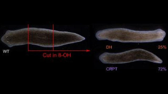
Flatworms treated with a drug inhibiting electrical synapses either repaired themselves back into their normal form (72 percent), into a two-headed form (25 percent), or did not regenerate (3 percent).

FALLON DURANT, ALLEN DISCOVERY CENTER AT TUFTS UNIVERSITY

Tufts University scientists demonstrated that they can permanently alter the resulting body shape of a regenerating flatworm by manipulating its synapses, according to a study published today (May 24) in Biophysical Journal.

Flatworms (Dugesia japonica) have a remarkable capacity for repairing themselves; if they’re sliced in half, they can reconstruct their deficient half to make themselves whole again, producing a duplicate of their pre-injury form.

Here, scientists used a drug to temporarily inhibit communication between flatworms’ electrical synapses, which differ from chemical synapses by using channel proteins to span small gaps between neurons. This molecular block made one quarter of the worms regenerate into a two-headed form ...

Interested in reading more?

Become a Member of

The Scientist Logo
Receive full access to more than 35 years of archives, as well as TS Digest, digital editions of The Scientist, feature stories, and much more!
Already a member? Login Here
Illustration of a developing fetus surrounded by a clear fluid with a subtle yellow tinge, representing amniotic fluid.
January 2026, Issue 1

What Is the Amniotic Fluid Composed of?

The liquid world of fetal development provides a rich source of nutrition and protection tailored to meet the needs of the growing fetus.

View this Issue
Skip the Wait for Protein Stability Data with Aunty

Skip the Wait for Protein Stability Data with Aunty

Unchained Labs
Graphic of three DNA helices in various colors

An Automated DNA-to-Data Framework for Production-Scale Sequencing

illumina
Exploring Cellular Organization with Spatial Proteomics

Exploring Cellular Organization with Spatial Proteomics

Abstract illustration of spheres with multiple layers, representing endoderm, ectoderm, and mesoderm derived organoids

Organoid Origins and How to Grow Them

Thermo Fisher Logo

Products

Brandtech Logo

BRANDTECH Scientific Introduces the Transferpette® pro Micropipette: A New Twist on Comfort and Control

Biotium Logo

Biotium Launches GlycoLiner™ Cell Surface Glycoprotein Labeling Kits for Rapid and Selective Cell Surface Imaging

Colorful abstract spiral dot pattern on a black background

Thermo Scientific X and S Series General Purpose Centrifuges

Thermo Fisher Logo
Abstract background with red and blue laser lights

VANTAstar Flexible microplate reader with simplified workflows

BMG LABTECH