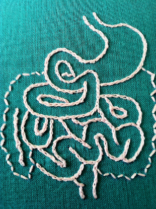
 FLICKR, HEY PAUL STUDIOSStarting with a sample of human intestine, researchers at Children’s Hospital Los Angeles and their colleagues have grown functioning segments of human intestine in the abdominal cavities of mice, according to a study published last week (January 8) in American Journal of Physiology - Gastrointestinal and Liver Physiology. The organs recapitulated the correct intestinal structure, with an epithelium surrounding a lumen and supporting mesenchyme and muscle, as well as tight junctions, microvilli, and other features of the human intestines. Moreover, the intestinal segments could absorb and break down complex sugars, demonstrating their ability to function like real intestines.
Last October, another group of researchers published in Nature Medicine their success using pluripotent stem cells to grow miniature human intestines inside the kidneys of immunosuppressed mice.
In addition to serving as a model for human intestinal function and dysfunction, with this latest achievement toward growing functional human intestines in mice, the researchers hope that the strategy could one day offer a treatment for intestinal failure, which accounts for 2 percent of neonatal intensive care unit admissions and kills nearly a third of affected babies. “Having a surrogate system in which you can prove the tissue grows properly is important,” Tracy Grikscheit of Children’s Hospital Los Angeles told New Scientist. “Every time you scale things up—from, say, a mouse to the size of a human ...