
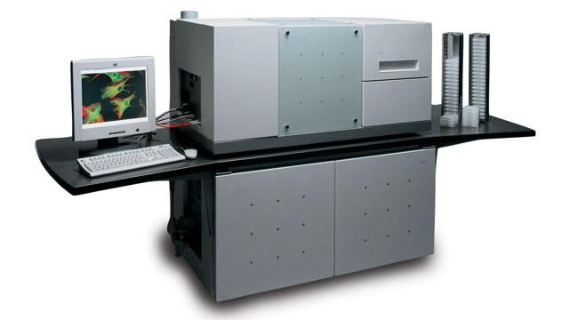
 THE OPERA: a high-content screening system from PerkinElmerCOURTESY OF PERKINELMER
Whether you’re measuring shape-shifting in a single dish of cells or treating a multiwell plate of them with thousands of different compounds to identify drug targets, analyzing a single readout of biological activity probably won’t give you the whole story. One way of assessing multiple features of a cell or small organism is to use high-content screening (HCS) microscope systems, which let you automate image acquisition and analysis. “This area has and is going to have a big effect on how we start to understand biology” following new genetics and genomics findings, says Ann Hoffman, a senior principal scientist in the imaging department of Roche Discovery Technologies at Hoffmann-La Roche in Nutley, New Jersey.
Dedicated HCS microscopes have been around for at least a decade. ...


