
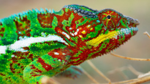
 NATURE COMMUNICATIONS, J. TEYSSIER ET AL. Chameleon colors aren’t just for camouflage. When panther chameleons (Furcifer pardalis) in Madagascar fight over territory, a dazzling display precedes their contests: resting males, typically green and inconspicuous, turn yellow or orange; red patches on their bodies can brighten, and blues can fade to whitish tints. The color changes, which are completely reversible and occur within minutes, are not the result of shifts in pigments alone. Results published today (March 10) in Nature Communications suggest they are the result of quick changes to light-reflecting guanine nanocrystals, which create structural color within chameleon skin.
Two layers of cells known as iridophores contain these nanocrystals. A superficial layer, known as S-iridophores, actively alters the spacing of these crystals to cause the rapid color changes, while a deeper layer, made up of D-iridophores, reflects a broader spectrum of light near the infrared wavelengths. In addition to camouflage and flashy fights, these cells may play a key part in keeping these lizards cool.
“People generally assume that color change in chameleons is well understood, and I don’t think it is at all,” said Randall Morrison of McDaniel College in Maryland who was not involved with the study. “This whole notion of the tunable photonic crystals is a new way to look at physiological color change in animals.”
Michel Milinkovitch of the University ...


