Image of the Day: Hippocampal Jalapeno

To tease apart brain regions involved in forming versus remembering memories, scientists engineered mice whose brain cells could be manipulated and tagged.

Written byThe Scientist
| 1 min read

Register for free to listen to this article
Listen with Speechify
0:00
1:00
Share

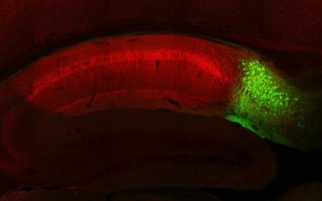
Fluorescently labeled cells in a mouse brain, with subiculum neurons in green and CA1 neurons in redRIKEN-MIT CENTER FOR NEURAL CIRCUIT GENETICS Researchers demonstrated that the mouse subiculum, a brain region associated with the hippocampus, is important for recalling certain types of memories, but it doesn’t appear to play a role in forming them. When they optogenetically turned off neurons within the subiculum, mice’s abilities to retrieve a memory they had previously formed was disrupted.

Some scientists think that brain circuits responsible for forming memories are the same as those necessary for retrieving them, write the authors in their report. These data, however, offer evidence to the contrary.

See D.S. Roy et al., “Distinct neural circuits for the formation and retrieval of episodic memories,” Cell, doi:10.1016/j.cell.2017.07.013, 2017.

Interested in reading more?

Become a Member of

The Scientist Logo
Receive full access to more than 35 years of archives, as well as TS Digest, digital editions of The Scientist, feature stories, and much more!
Already a member? Login Here
Illustration of a developing fetus surrounded by a clear fluid with a subtle yellow tinge, representing amniotic fluid.
January 2026, Issue 1

What Is the Amniotic Fluid Composed of?

The liquid world of fetal development provides a rich source of nutrition and protection tailored to meet the needs of the growing fetus.

View this Issue
Skip the Wait for Protein Stability Data with Aunty

Skip the Wait for Protein Stability Data with Aunty

Unchained Labs
Graphic of three DNA helices in various colors

An Automated DNA-to-Data Framework for Production-Scale Sequencing

illumina
Exploring Cellular Organization with Spatial Proteomics

Exploring Cellular Organization with Spatial Proteomics

Abstract illustration of spheres with multiple layers, representing endoderm, ectoderm, and mesoderm derived organoids

Organoid Origins and How to Grow Them

Thermo Fisher Logo

Products

Brandtech Logo

BRANDTECH Scientific Introduces the Transferpette® pro Micropipette: A New Twist on Comfort and Control

Biotium Logo

Biotium Launches GlycoLiner™ Cell Surface Glycoprotein Labeling Kits for Rapid and Selective Cell Surface Imaging

Colorful abstract spiral dot pattern on a black background

Thermo Scientific X and S Series General Purpose Centrifuges

Thermo Fisher Logo
Abstract background with red and blue laser lights

VANTAstar Flexible microplate reader with simplified workflows

BMG LABTECH