Image of the Day: Tardigrade Eggs

The studded cells belong to a newly discovered species of water bear, Macrobiotus shonaicus.

Written byThe Scientist
| 1 min read

Register for free to listen to this article
Listen with Speechify
0:00
1:00
Share

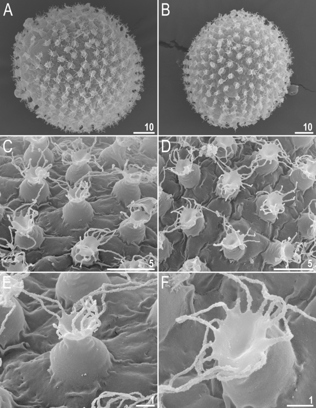
Scanning electron micrograph of tardigrade (Macrobiotus shonaicus) eggs. A-B: entire eggs with flexible filaments on the egg processes; C-D: egg processes with filaments; E-F: close-up of a single egg process. All scale bars in micrometersSTEC ET AL.

Researchers announced the discovery of a new species of tardigrade in PLOS ONE on Wednesday (February 28). The animal is named Macrobiotus shonaicus for the Shōnai region in Japan where it was collected. Study coauthor Kazaharu Arakawa, a molecular biologist at Keio University, collected the critter in a clump of moss he scraped from the surface of his apartment complex’s parking lot.

The new species was added to the hufelandi group of tardigrades, which was previously represented by just one other species, M. hufelandi. Hufelandi tardigrades’ eggs have similar characteristics, notably, structures on their surface called egg processes. M. shonaicus has thin, flexible filaments on the ends of the egg processes that most likely enhance the egg’s adhesiveness.

D. Stec et al., “An integrative description ...

Interested in reading more?

Become a Member of

The Scientist Logo
Receive full access to more than 35 years of archives, as well as TS Digest, digital editions of The Scientist, feature stories, and much more!
Already a member? Login Here
Illustration of a developing fetus surrounded by a clear fluid with a subtle yellow tinge, representing amniotic fluid.
January 2026, Issue 1

What Is the Amniotic Fluid Composed of?

The liquid world of fetal development provides a rich source of nutrition and protection tailored to meet the needs of the growing fetus.

View this Issue
Skip the Wait for Protein Stability Data with Aunty

Skip the Wait for Protein Stability Data with Aunty

Unchained Labs
Graphic of three DNA helices in various colors

An Automated DNA-to-Data Framework for Production-Scale Sequencing

illumina
Exploring Cellular Organization with Spatial Proteomics

Exploring Cellular Organization with Spatial Proteomics

Abstract illustration of spheres with multiple layers, representing endoderm, ectoderm, and mesoderm derived organoids

Organoid Origins and How to Grow Them

Thermo Fisher Logo

Products

Brandtech Logo

BRANDTECH Scientific Introduces the Transferpette® pro Micropipette: A New Twist on Comfort and Control

Biotium Logo

Biotium Launches GlycoLiner™ Cell Surface Glycoprotein Labeling Kits for Rapid and Selective Cell Surface Imaging

Colorful abstract spiral dot pattern on a black background

Thermo Scientific X and S Series General Purpose Centrifuges

Thermo Fisher Logo
Abstract background with red and blue laser lights

VANTAstar Flexible microplate reader with simplified workflows

BMG LABTECH