In the 1860s, physician Hermann von Helmholtz did a simple experiment to understand how the world stays still during eye movements. With a still head, he closed one eye and swiveled the other to look around. Despite the rapid darting of the open eye, he noticed the image of the surroundings appeared stable rather than blurred. Next, instead of moving the eye naturally, he gently pushed it around in the socket with a finger and noticed the chaotic movement of the view. Why does the world shift when an external force is used to move the eye, but not when it swivels on its own?
Von Helmholtz proposed that when an animal decides to move its eyes naturally, certain areas of the brain receive a duplicate of this command called the efference copy, which signals that the upcoming motion of the world is a result of eye movement and not actual shifting of the environment.1 This message is absent when an external force moves the visual field. “When the finger hijacks the movement, it takes away the efference copy and we see what happens in a world without one,” said Tomas Vega-Zuniga, a neuroscientist at the Institute of Science and Technology Austria (ISTA). The efference copy plays a crucial role in an animal’s ability to differentiate its own motion from that of the surrounding world, which in turn is essential for coherent perception and behavior.2
Understanding the neural mechanisms of this brain-body coordination has intrigued neuroscientists for decades. Previous investigations have suggested that the efference copy originates in various thalamic and cortical regions of the brain. Now, researchers at ISTA have shown that a region of the thalamus, called the ventral lateral geniculate nucleus (vLGN), serves as an interface between visual and motor neural circuits and is responsible for correcting self-motion-induced blur. These findings in mice, published in Nature Neuroscience, could help researchers better understand how an organism’s senses faithfully represent the world and enable appropriate behavior.3
Visual perception is complex and requires seamless communication between different brain areas. One such region that integrates visual and other sensory perceptions with movement is the superior colliculus.4 This multi-layered structure receives visual information directly from the retina, as well as indirectly via the visual cortex. “The superior colliculus is like a map of the world. It knows where things are in space,” said Vega-Zuniga, a study coauthor. In previous experiments in primates, researchers showed that the superior colliculus sends signals to areas in the cortex that control eye movements. So, Vega-Zuniga and his colleagues hypothesized that neurons that send information to the superior colliculus could serve as a source of the efference copy and correct motion-induced blur.
An important function of the efference copy is to block certain sensory inputs to maintain coherent perception for the organism. For example, auditory signaling in crickets is diminished during their own chirps, so as not to desensitize hearing at other times. Since this is achieved through suppression or inhibition, the team focused on the vLGN, which forms inhibitory connections with neurons in superior colliculus.

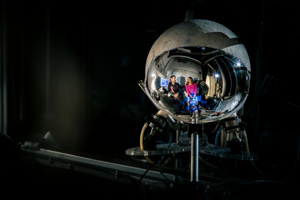
Neuroscientists Olga Symonova and Tomas Vega-Zuniga, along with their colleagues, used a custom-built imaging setup to visualize mice brain activity while the animals were awake and behaving.
Institute of Science and Technology Austria
Vega-Zuniga and his colleagues wanted to determine how the vLGN modulates the activity of superior colliculus neurons in a mouse that is awake and performing diverse behaviors. They built an experimental setup in which the animal could run freely on a tiny ball while they peered into its brain through a tiny window in its skull. Using calcium imaging to visualize the activity of the vLGN neurons, the researchers observed that the thalamic neurons responded to both visual stimuli and movement, including eye movement, making them well-suited to modulate visual signals in the superior colliculus in response to self-motion.
However, the question remained: Does the vLGN produce an efference copy that helps to differentiate between self and external motion? The researchers recorded vLGN neuronal responses to natural eye movement or eye movement simulated by the movement of a virtual environment around the mice, representing self and external motion, respectively. They found that the vLGN neurons only responded to self-motion. Additionally, when the team blocked vLGN activity, the neuronal responses to eye movement in the superior colliculus became longer and more frequent, suggesting that the vLGN shortens the effective time of visual exposure during movement, thus reducing blur.
To confirm this, Vega-Zuniga and his colleagues tested how well the mice could perceive depth, which not only requires both visual and movement perception, but is detrimentally affected by blurry vision. They observed that mice in which the vLGN output was blocked showed reduced avoidance of a cliff in the behavioral arena, demonstrating difficulty in judging depth. Based on this, the authors suggested that the vLGN is important for visual perception during self-generated movements.
However, some researchers think it’s too early to infer this with conviction. “They need to demonstrate that there’s an informative gate at the level of the vLGN that tells the cortex, ‘Look, it's not the external world that is moving, it's you that is moving,’” said Maria Morrone, a neuroscientist at the University of Pisa, who was not involved in the study. “That will change the field of active vision.”
Aman Saleem, a behavioral neuroscientist at University College London who did not contribute to the study, expressed similar reservations. “Does [the vLGN] have a modulatory influence or is it actively encoding information?” he questioned. While he appreciates the in-depth characterization of the circuits and behaviors related to the vLGN, he is looking forward to data on the detailed computations at each step, from the information coming through the retina, to what the animal sees when it makes the movement.
- Sun LD, Goldberg ME. Corollary discharge and oculomotor proprioception: Cortical mechanisms for spatially accurate vision.Annu Rev Vis Sci. 2016;2:61-84.
- Crapse TB, Sommer MA. Corollary discharge across the animal kingdom.Nat Rev Neurosci. 2008;9(8):587-600.
- Vega-Zuniga T, et al. A thalamic hub-and-spoke network enables visual perception during action by coordinating visuomotor dynamics.Nat Neurosci. 2025;28:627-639.
- Massot C, et al. Sensorimotor transformation elicits systematic patterns of activity along the dorsoventral extent of the superior colliculus in the macaque monkey.Commun Biol. 2019;2(1):287.


