

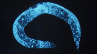
 C. elegansWIKIMEDIA, NATIONAL HUMAN GENOME RESEARCH INSTITUTEResearchers from the United States and Vienna have simultaneously imaged the activity of all 302 neurons of C. elegans, revealing how quickly nervous impulses traverse the worm. The team then used the technique, which involves engineering neurons to light up when firing, to image the brains of transparent zebrafish larvae. While scientists have long mapped the neuronal connections of various organisms—the entire connectome of C. elegans was first published in 1986, for example—the new results, published yesterday (May 18) in Nature Methods, represent the first time that the activity of an organism’s entire nervous system has been viewed in real time.
“The connectome and activity are entirely complementary,” Misha Ahrens, a neurobiologist at the Howard Hughes Medical Institute's Janelia Farm Research Campus in Ashburn, Virginia, told Nature News. “You’re not going to understand the nervous system by observing one or the other.”
“What’s very impressive about it is that it is such an elegantly simple implementation,” Aravinthan Samuel, a professor of physics at Harvard University who was not part of the research team, said in a press release. “I could imagine many labs adopting this.”
To achieve this imaging success, neuroscientist Alipasha Vaziri of the University of Vienna and his colleagues turned to light-field deconvolution microscopy, which yields 3–D images based on images from a set of tiny lenses. With up to 50 images per second, ...


