Next Generation: A Molecular Camera

Knocking electrons out of atomic orbit with a laser allows researchers to take femtosecond-scale “movies” of molecules in motion.

Written byHannah Waters
| 3 min read

Register for free to listen to this article
Listen with Speechify
0:00
3:00
Share

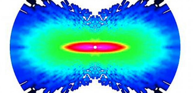
A nitrogen molecule shifts during the time between laser pulses, one femtosecond. The atoms' movements are shown as a measure of increasing angular momentum, on a scale from dark blue to pink, with pink showing the region of greatest momentum. IMAGE COURTESY OF COSMIN BLAGA

THE DEVICE: Molecules are in constant motion as electrons swarm around inside atoms, slightly flexing the atomic bonds and shifting the molecule’s shape. Using standard tools found in physical chemistry labs, physicist Cosmin Blaga from Ohio State University and his colleagues have executed a technique to take rapid snapshots of molecules in motion, published last week (March 7) in Nature.

By shooting a laser through a tiny hole into a vacuum chamber, Blaga created an infrared laser field. The laser’s intensity knocks an electron out of atomic orbit just for a few femtoseconds, or 10-15 seconds. In that short time before the electron is pulled back into orbit, the bonds holding the molecule’s atoms stretch. And, even if they shift by only a mere angstrom ...

Interested in reading more?

Become a Member of

The Scientist Logo
Receive full access to more than 35 years of archives, as well as TS Digest, digital editions of The Scientist, feature stories, and much more!
Already a member? Login Here
Illustration of a developing fetus surrounded by a clear fluid with a subtle yellow tinge, representing amniotic fluid.
January 2026, Issue 1

What Is the Amniotic Fluid Composed of?

The liquid world of fetal development provides a rich source of nutrition and protection tailored to meet the needs of the growing fetus.

View this Issue
Skip the Wait for Protein Stability Data with Aunty

Skip the Wait for Protein Stability Data with Aunty

Unchained Labs
Graphic of three DNA helices in various colors

An Automated DNA-to-Data Framework for Production-Scale Sequencing

illumina
Exploring Cellular Organization with Spatial Proteomics

Exploring Cellular Organization with Spatial Proteomics

Abstract illustration of spheres with multiple layers, representing endoderm, ectoderm, and mesoderm derived organoids

Organoid Origins and How to Grow Them

Thermo Fisher Logo

Products

Brandtech Logo

BRANDTECH Scientific Introduces the Transferpette® pro Micropipette: A New Twist on Comfort and Control

Biotium Logo

Biotium Launches GlycoLiner™ Cell Surface Glycoprotein Labeling Kits for Rapid and Selective Cell Surface Imaging

Colorful abstract spiral dot pattern on a black background

Thermo Scientific X and S Series General Purpose Centrifuges

Thermo Fisher Logo
Abstract background with red and blue laser lights

VANTAstar Flexible microplate reader with simplified workflows

BMG LABTECH