Potential Route for Zika from Mother to Fetus

Placental macrophages called Hofbauer cells are particularly vulnerable to infection by the virus, a study shows.

Written byCatherine Offord
| 2 min read

Register for free to listen to this article
Listen with Speechify
0:00
2:00
Share

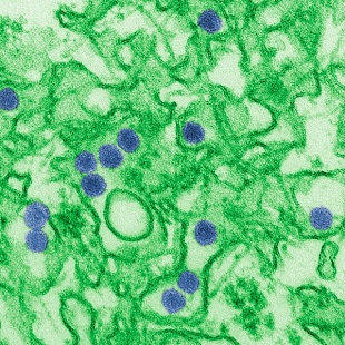
Zika virus particles (blue) WIKIMEDIA, CDC, CYNTHIA GOLDSMITHThe Zika virus may pass from a pregnant woman to her developing fetus via placental immune cells known as Hofbauer cells, according to a study published last week (May 27) in Cell Host & Microbe. The findings highlight a possible transmission route for the virus, which has been linked to microcephaly and other birth defects in more than a quarter of babies born to infected mothers.

“Our study indicates that this cell type may be a target for Zika virus in the placenta,” study coauthor Rana Chakraborty of Emory University School of Medicine said in a statement. “Replication in these cells may allow the virus to cross the placental barrier and enter the fetal circulation.”

Until now, the path of the virus from mother to fetus has remained elusive. In fact, some cells in the outermost cell layer of the placenta have been shown to be fairly resistant to Zika infection and may even act as a barrier to transmission.

In the present study, the team used human placentae from five donors to identify Hofbauer cells as particularly vulnerable to Zika infection. Cells called cytotrophoblasts present in the middle layer of ...

Interested in reading more?

Become a Member of

The Scientist Logo
Receive full access to more than 35 years of archives, as well as TS Digest, digital editions of The Scientist, feature stories, and much more!
Already a member? Login Here

Related Topics

Meet the Author

  • After undergraduate research with spiders at the University of Oxford and graduate research with ants at Princeton University, Catherine left arthropods and academia to become a science journalist. She has worked in various guises at The Scientist since 2016. As Senior Editor, she wrote articles for the online and print publications, and edited the magazine’s Notebook, Careers, and Bio Business sections. She reports on subjects ranging from cellular and molecular biology to research misconduct and science policy. Find more of her work at her website.

    View Full Profile
Share
Illustration of a developing fetus surrounded by a clear fluid with a subtle yellow tinge, representing amniotic fluid.
January 2026, Issue 1

What Is the Amniotic Fluid Composed of?

The liquid world of fetal development provides a rich source of nutrition and protection tailored to meet the needs of the growing fetus.

View this Issue
Skip the Wait for Protein Stability Data with Aunty

Skip the Wait for Protein Stability Data with Aunty

Unchained Labs
Graphic of three DNA helices in various colors

An Automated DNA-to-Data Framework for Production-Scale Sequencing

illumina
Exploring Cellular Organization with Spatial Proteomics

Exploring Cellular Organization with Spatial Proteomics

Abstract illustration of spheres with multiple layers, representing endoderm, ectoderm, and mesoderm derived organoids

Organoid Origins and How to Grow Them

Thermo Fisher Logo

Products

Brandtech Logo

BRANDTECH Scientific Introduces the Transferpette® pro Micropipette: A New Twist on Comfort and Control

Biotium Logo

Biotium Launches GlycoLiner™ Cell Surface Glycoprotein Labeling Kits for Rapid and Selective Cell Surface Imaging

Colorful abstract spiral dot pattern on a black background

Thermo Scientific X and S Series General Purpose Centrifuges

Thermo Fisher Logo
Abstract background with red and blue laser lights

VANTAstar Flexible microplate reader with simplified workflows

BMG LABTECH