

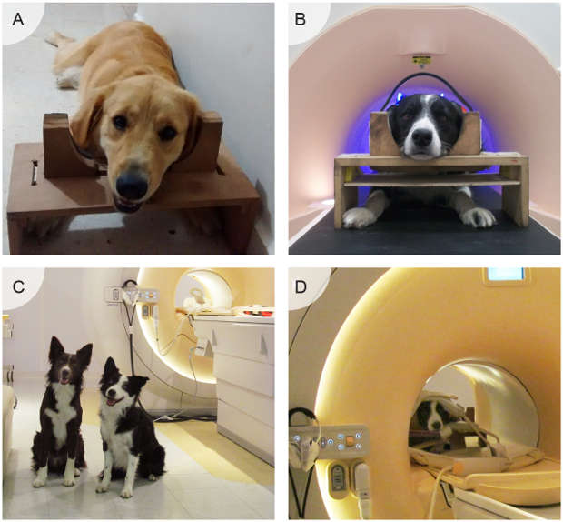
 L.V. CUAYA ET AL., PLOS ONEResearchers from the National Autonomous University of Mexico have studied the brains of seven dogs trained to lie down but stay attentive inside an MRI scanner. The team presented the animals with photos of human faces or of objects. The object images elicited very little brain activity, but the human pictures triggered a burst of activity: the bilateral temporal cortex lit up the most, and the team saw additional activity in the frontal cortex, the caudate nucleus, and the thalamus.
“This portion of the temporal cortex in dogs could be anatomically and functionally similar to regions found in other species, like humans, non-human primates and sheep, which suggests a high degree of evolutionary conservation of the ventral visual pathway for face processing,” the researchers wrote in their paper, published this week (March 2) in PLOS ONE.
Studies of how dogs recognize human faces, then, may inform our understanding of human facial perception. A study from February 2015, for example, tested dogs’ ability to identify emotional expressions when shown pictures of just one half a human face, and the canines performed well.
“The recognition of human faces by dogs could be an essential factor for establishing attachment with humans,” the authors of the new PLOS ONE ...


