Reprogrammed Glia Improve Symptoms in a Mouse Model of Parkinson’s

By converting glial cells into dopaminergic neurons, scientists were able to partially rescue motor behavior in mice.

Written byDiana Kwon
| 3 min read

Register for free to listen to this article
Listen with Speechify
0:00
3:00
Share

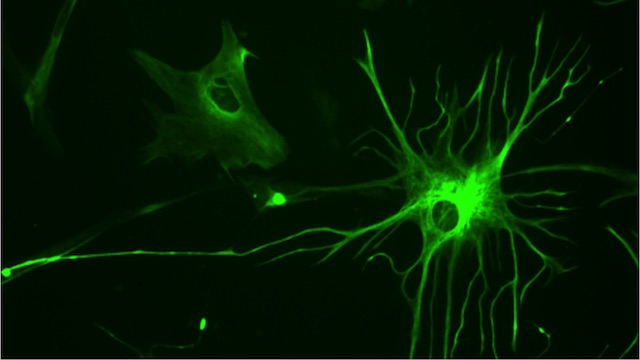
A human astrocyteWIKIMEDIA, BRUNO PASCAL

Parkinson’s, a neurodegenerative disease that primarily affects the motor system, is marked by a progressive loss of dopaminergic neurons in the brain. While current treatments are aimed at replenishing dopamine levels, none are able to restore the lost cells. Now, scientists have devised a way to reprogram glial cells into active dopamine neurons that can partially restore motor function in a mouse model of Parkinson’s. This proof-of-principle study could pave the way to a new treatment for the disease, researchers reported today (April 10) in Nature Biotechnology.

“In Parkinson’s disease, dopamine neurons die, but at the same time, because of inflammation . . . some glial cells become reactive and proliferate,” said coauthor Ernest Arenas, a molecular neurobiologist at the Karolinska Institute in Sweden. “So ...

Interested in reading more?

Become a Member of

The Scientist Logo
Receive full access to more than 35 years of archives, as well as TS Digest, digital editions of The Scientist, feature stories, and much more!
Already a member? Login Here

Related Topics

Meet the Author

  • Diana is a freelance science journalist who covers the life sciences, health, and academic life. She’s a regular contributor to The Scientist and her work has appeared in several other publications, including Scientific American, Knowable, and Quanta. Diana was a former intern at The Scientist and she holds a master’s degree in neuroscience from McGill University. She’s currently based in Berlin, Germany.

    View Full Profile
Share
Illustration of a developing fetus surrounded by a clear fluid with a subtle yellow tinge, representing amniotic fluid.
January 2026, Issue 1

What Is the Amniotic Fluid Composed of?

The liquid world of fetal development provides a rich source of nutrition and protection tailored to meet the needs of the growing fetus.

View this Issue
Skip the Wait for Protein Stability Data with Aunty

Skip the Wait for Protein Stability Data with Aunty

Unchained Labs
Graphic of three DNA helices in various colors

An Automated DNA-to-Data Framework for Production-Scale Sequencing

illumina
Exploring Cellular Organization with Spatial Proteomics

Exploring Cellular Organization with Spatial Proteomics

Abstract illustration of spheres with multiple layers, representing endoderm, ectoderm, and mesoderm derived organoids

Organoid Origins and How to Grow Them

Thermo Fisher Logo

Products

Brandtech Logo

BRANDTECH Scientific Introduces the Transferpette® pro Micropipette: A New Twist on Comfort and Control

Biotium Logo

Biotium Launches GlycoLiner™ Cell Surface Glycoprotein Labeling Kits for Rapid and Selective Cell Surface Imaging

Colorful abstract spiral dot pattern on a black background

Thermo Scientific X and S Series General Purpose Centrifuges

Thermo Fisher Logo
Abstract background with red and blue laser lights

VANTAstar Flexible microplate reader with simplified workflows

BMG LABTECH