
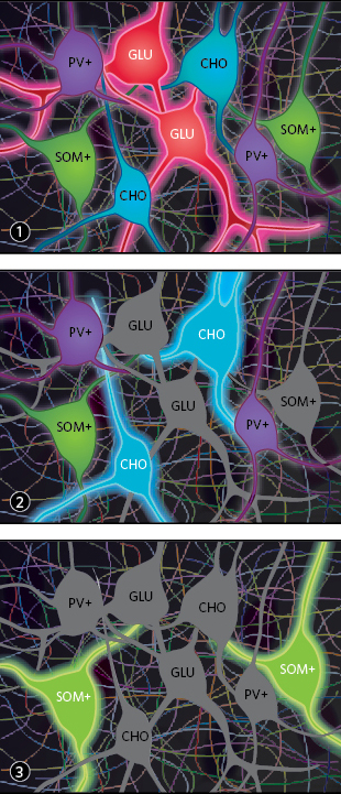
 INTERWEBS: Activating glutamatergic (GLU) neurons optogenetically excites all other labeled cells types in the basal forebrain of a mouse (1). When cholinergic (CHO) neurons are turned on, they inhibit GLU neurons and excite a subtype of GABAergic neurons containing parvalbumin (PV+). They also excite and inhibit somatostatin-containing (SOM+) GABAergic neurons (2). SOM+ neurons, the only cell type less active during REM sleep and waking, inhibit other neuron types (3). PV+ neurons have minimal or no effects on the other cell types (not shown).© KIMBERLY BATTISTAThe paper
M. Xu et al., “Basal forebrain circuit for sleep-wake control,” Nature Neuroscience, 18:1641-47, 2015.
Early studies attempting to untangle the neurological basis of sleep typically removed or injured part of an animal’s brain to measure the effects. The results implicated a region called the basal forebrain in inducing sleep, yet some studies indicated that it was important for arousal. “The impression is that maybe in that region there’s a mixture of mechanisms,” says Yang Dan, a neurobiologist at the University of California, Berkeley. “But that’s not a very satisfactory answer.”
Dan sought to identify which cells in the basal forebrain promote which brain state. The region contains three main types of neurons: cholinergic, glutamatergic, and GABAergic. Dan and her colleagues further classified the GABAergic neurons into those containing somatostatin (SOM+) or parvalbumin (PV+).
The researchers optogenetically activated each of these four cell types in mice to locate them and track their activity. The cholinergic, glutamatergic, and PV+ GABAergic neurons typically fired multiple times per second when the mice were awake or in REM sleep, but less often during non-REM sleep, a sleep stage in which the brain is less aroused overall. In contrast, non-REM sleep was when the SOM+ GABAergic neurons were most active.
Dan’s team then fired a laser pulse to stimulate the different ...


