SSRI Consumption During Pregnancy Linked to Changes in Babies’ Brains

MRI data suggest prenatal exposure to the medication increases the volume of brain regions vital to processing emotions.

Written byAshley Yeager
| 2 min read

Register for free to listen to this article
Listen with Speechify
0:00
2:00
Share

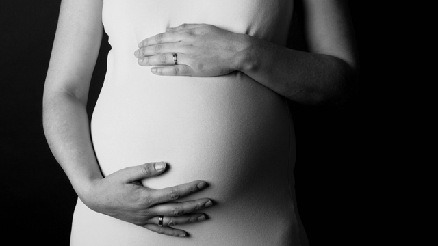
FLICKR, TATIANA DVPMothers who take selective serotonin reuptake inhibitors (SSRIs), a class of commonly used antidepressants, while pregnant have babies with distinct structural changes to their brains, researchers report today (April 9) in JAMA Pediatrics. MRI scans of the babies’ brains revealed exposure to the drugs in the womb increased the volumes of the babies’ amygdalae and insular cortices—regions that play a role in processing emotions.

“Hopefully these results highlight the fact that something could be going on here,” study coauthor Claudia Lugo-Candelas, a postdoctoral research fellow at Columbia University tells Time. “They point to the fact that there is a signal—we don’t know what it means, or don’t know how long it might last. But we know it’s worth studying.”

The number of women using SSRIs while pregnant is increasing, but not much is known about how the medication might affect the brains of developing babies. Studies in animals suggest exposure to SSRIs can change the offspring’s brain circuitry and lead to depressive-like behaviors and anxiety later in life.

In the new study, Lugo-Candelas and her colleagues studied the brains of 98 human infants, 16 babies whose mothers were treated ...

Interested in reading more?

Become a Member of

The Scientist Logo
Receive full access to more than 35 years of archives, as well as TS Digest, digital editions of The Scientist, feature stories, and much more!
Already a member? Login Here

Related Topics

Meet the Author

  • Ashley started at The Scientist in 2018. Before joining the staff, she worked as a freelance editor and writer, a writer at the Simons Foundation, and a web producer at Science News, among other positions. She holds a bachelor’s degree in journalism from the University of Tennessee, Knoxville, and a master’s degree in science writing from MIT. Ashley edits the Scientist to Watch and Profile sections of the magazine and writes news, features, and other stories for both online and print.

    View Full Profile
Share
Illustration of a developing fetus surrounded by a clear fluid with a subtle yellow tinge, representing amniotic fluid.
January 2026, Issue 1

What Is the Amniotic Fluid Composed of?

The liquid world of fetal development provides a rich source of nutrition and protection tailored to meet the needs of the growing fetus.

View this Issue
Skip the Wait for Protein Stability Data with Aunty

Skip the Wait for Protein Stability Data with Aunty

Unchained Labs
Graphic of three DNA helices in various colors

An Automated DNA-to-Data Framework for Production-Scale Sequencing

illumina
Exploring Cellular Organization with Spatial Proteomics

Exploring Cellular Organization with Spatial Proteomics

Abstract illustration of spheres with multiple layers, representing endoderm, ectoderm, and mesoderm derived organoids

Organoid Origins and How to Grow Them

Thermo Fisher Logo

Products

Brandtech Logo

BRANDTECH Scientific Introduces the Transferpette® pro Micropipette: A New Twist on Comfort and Control

Biotium Logo

Biotium Launches GlycoLiner™ Cell Surface Glycoprotein Labeling Kits for Rapid and Selective Cell Surface Imaging

Colorful abstract spiral dot pattern on a black background

Thermo Scientific X and S Series General Purpose Centrifuges

Thermo Fisher Logo
Abstract background with red and blue laser lights

VANTAstar Flexible microplate reader with simplified workflows

BMG LABTECH