Stems Cells Ushered into Embryonic Development

The right mix of mouse embryonic stem cells in a dish will start forming early embryonic patterns, according to two studies.

Written byKerry Grens
| 1 min read

Register for free to listen to this article
Listen with Speechify
0:00
1:00
Share

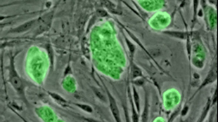
WIKIMEDIA, NSFAlthough embryonic stem cells can form any tissue type in vitro, they have always given researchers trouble when it came to developing a properly organized embryo. Alfonso Martinez-Arias at Cambridge University and colleagues reported in the November issue of Development that if aggregated in the right numbers—roughly 300— embryonic stem cells will begin to arrange themselves in patterns that mimic early embryonic development.

“Much to our surprise, after three days in culture they begin a series [of] movements that resemble those that organize the embryo and acquire coordinates and, as in the embryo, the seeds of the different tissues,” Martinez-Arias told Motherboard in an e-mail.

According to a statement, the researchers could then direct cell fate. “In one of the experiments, for example, activation of a particular signal at the correct time elicits the appearance of the mesoderm, endoderm and ectoderm—the precursors of all cell types—with a spatial organization similar to that of an embryo.”

In a second paper in Development, the researchers used their experimental design to explore the development of the neuromesodermal precursor, which will go on to form the spinal cord.

Interested in reading more?

Become a Member of

The Scientist Logo
Receive full access to more than 35 years of archives, as well as TS Digest, digital editions of The Scientist, feature stories, and much more!
Already a member? Login Here

Related Topics

Meet the Author

  • kerry grens

    Kerry served as The Scientist’s news director until 2021. Before joining The Scientist in 2013, she was a stringer for Reuters Health, the senior health and science reporter at WHYY in Philadelphia, and the health and science reporter at New Hampshire Public Radio. Kerry got her start in journalism as a AAAS Mass Media fellow at KUNC in Colorado. She has a master’s in biological sciences from Stanford University and a biology degree from Loyola University Chicago.

    View Full Profile
Share
Illustration of a developing fetus surrounded by a clear fluid with a subtle yellow tinge, representing amniotic fluid.
January 2026, Issue 1

What Is the Amniotic Fluid Composed of?

The liquid world of fetal development provides a rich source of nutrition and protection tailored to meet the needs of the growing fetus.

View this Issue
Redefining Immunology Through Advanced Technologies

Redefining Immunology Through Advanced Technologies

Skip the Wait for Protein Stability Data with Aunty

Skip the Wait for Protein Stability Data with Aunty

Unchained Labs
Graphic of three DNA helices in various colors

An Automated DNA-to-Data Framework for Production-Scale Sequencing

illumina
Exploring Cellular Organization with Spatial Proteomics

Exploring Cellular Organization with Spatial Proteomics

Products

nuclera logo

Nuclera eProtein Discovery System installed at leading Universities in Taiwan

Brandtech Logo

BRANDTECH Scientific Introduces the Transferpette® pro Micropipette: A New Twist on Comfort and Control

Biotium Logo

Biotium Launches GlycoLiner™ Cell Surface Glycoprotein Labeling Kits for Rapid and Selective Cell Surface Imaging

Colorful abstract spiral dot pattern on a black background

Thermo Scientific X and S Series General Purpose Centrifuges

Thermo Fisher Logo