

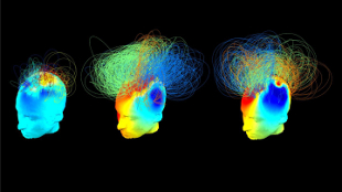
 Illustrated brain networks in two vegetative patients (left and middle), but one of whom imagined playing tennis (middle), alongside a healthy adult (right).SRIVAS CHENNUUsing electroencephalography (EEG)—a far simpler and cheaper brain imaging technique that functional magnetic resonance imaging (fMRI)—to compare brain activity of healthy adults with 32 vegetative or minimally conscious patients, University of Cambridge researchers found that “the rich and diversely connected networks that support awareness are characteristically impaired in patients, lacking the ability to efficiently integrate information across disparate regions via well-connected hubs,” they wrote in a PLOS Computational Biology paper published last week (October 16). Importantly, the researchers identified some patients with networks that were much more similar to those of healthy adults, and these patients showed signs of “hidden awareness,” such as being able to follow commands by imaging different activities.
“Being able to detect the recovery of brain networks in patients, alongside or even before they show behavioural signs of improvement, is very promising,” the authors said in a press release. “However, further work is essential to translate these scientific advances into viable tools that can be reliably used at the patients’ bedsides to accurately inform and guide their clinical care.”
Understanding consciousness has become a popular area of neuroscience research, as scientists aim to characterize brain activity patterns that can identify signs of awareness in seemingly vegetative patients. “[F]or patients diagnosed as vegetative and minimally conscious, and their families, this is far more than just an academic question—it takes on a very real significance,” Cambridge’s Srivas Chennu said in a statement. “Our research could ...


