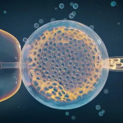
Simplified illustration of a scanning electron microscope and a transmission electron microscope.
| 2 min read
Both SEM and TEM reveal intricate details of samples, but they differ in how they generate an image.

electron microscopy

An illustration of a blue brain with the cerebellum highlighted in pink.

New Cilia Disassembly Pathway Revealed in Maturing Neurons

Jotham Austin, the advanced electron microscopy core director at the University of Chicago, stands in the facility beside an EM instrument, helping a student who is sitting at the computer.

Career Chat: Choosing a Core Career Track

JFT1 has reacted with hydrogen peroxide and APEX2 to create red fluorescence near APEX2 that remains separate from the green mitochondria.

FLEXing a Bright New Idea

A person’s hand reaching for wheat.

Close Encounters of Skin and Nerve Cells

Charting a New Course Through the Injured Brain

Using Mass Photometry to Assess and Characterize Protein Samples 

Rapidly Characterizing Complex Samples With Mass Photometry

Infographic detailing two volume electron microscopy modalities.

Infographic: Drivers of the Expansion of Volume Electron Microscopy

This image depicts the fruit fly nerve cord connectome. It highlights 930 neurons, a subset of the full set of reconstructed neurons.

The Expansion of Volume Electron Microscopy

 Dive into Cryo-EM’s History, Milestones, and Insights.

Cryo-EM: Building on a History of Invention and Innovation

Neurons in all sorts of different colors, some glowing

How Fear Restructures the Mouse Brain

Form Determines Function: Insights from Structural Biology

A scanning electron micrograph of a coculture of E. coli and Acinetobacter baylyi. Nanotubes can be seen extending from the E. coli.

What’s the Deal with Bacterial Nanotubes?

An illustration of a flask of bacteria, a weighted microscope slide, and two bacteria exchanging materials via nanotubes.

Infographic: Sources of Variation in Bacterial Nanotube Studies

Transforming Virology Research with Cryo-EM

Coronavirus Closeup, 1964

Coronavirus-Infected Cells Grow Filopodia

Image of the Day: Coronavirus Under the Scope

Imaging Chromatin to Deduce Function from Form

Aaron Klug, Developer of Crystallographic Electron Microscopy, Dies

Image of the Day: Feather Mites

Trending

Image of Atul Butte.

Biomedical Informatician Atul Butte Dies at 55

An illustration showing the accumulation of amyloid plaques between nerve cells, which is the underlying cause of disorders like Alzheimer’s and Parkinson’s disease.

A Gut Pathogen’s Unexpected Weapon Against Amyloid Diseases

Photo of John Calhoun crouches within his rodent utopia-turned-dystopia

Universe 25 Experiment

A group of newborn mice with their mother.

Maternal Iron Deficiency Can Trigger Sex Reversal in Mouse Embryos

Multimedia

Conceptual 3D illustration of mycoplasma colonies

Taking Control of Cellular Contamination with PCR Testing

sartorius logo
Digital microscopy image of cells in culture

Electronic Records in Cell Culture Management

Image of small blue creatures called Nergals. Some have hearts above their heads, which signify friendship. There is one Nergal who is sneezing and losing health, which is denoted by minus one signs floating around it.
June 2025, Issue 1

Nergal Networks: Where Friendship Meets Infection

A citizen science game explores how social choices and networks can influence how an illness moves through a population.

View this Issue
An illustration of green lentiviral particles.

Maximizing Lentivirus Recovery

cytiva logo
Unraveling Complex Biology with Advanced Multiomics Technology

Unraveling Complex Biology with Five-Dimensional Multiomics

Element Bioscience Logo
Resurrecting Plant Defense Mechanisms to Avoid Crop Pathogens

Resurrecting Plant Defense Mechanisms to Avoid Crop Pathogens

Twist Bio 
The Scientist Placeholder Image

Seeing and Sorting with Confidence

BD

Products

The Scientist Placeholder Image

Waters Enhances Alliance iS HPLC System Software, Setting a New Standard for End-to-End Traceability and Data Integrity 

The Scientist Placeholder Image

Agilent Unveils the Next Generation in LC-Mass Detection: The InfinityLab Pro iQ Series

agilent-logo

Agilent Announces the Enhanced 8850 Gas Chromatograph

parse-biosciences-logo

Pioneering Cancer Plasticity Atlas will help Predict Response to Cancer Therapies