Tasmanian Devils Developing Resistance to Transmissible Cancer

The marsupials’ genomes show evidence of a rapid evolutionary response to selection imposed by devil facial tumor disease.

Written byAbby Olena, PhD
| 3 min read

Register for free to listen to this article
Listen with Speechify
0:00
3:00
Share

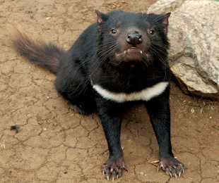
WIKIMEDIA, KERESHDuring the last 20 years, a contagious cancer has decimated Tasmanian devil (Sarcophilus harrisii) populations. Cancer cells, which are spread by biting, grow deadly tumors on the faces and mouths of the aggressive marsupials. Because devil facial tumor disease (DFTD) has been observed in almost all known populations and is nearly 100 percent fatal, epidemiological models have suggested that the most long-infected populations are facing extinction.

“But they’re currently surviving,” said Andrew Storfer of Washington State University. Now, he and his colleagues have the start of an explanation as to why. In a study published today (August 30) in Nature Communications, Storfer and an international team of researchers reported genomic evidence to suggest that Tasmanian devils are evolving resistance to DFTD.

“It’s such an important finding,” said Beata Ujvari of Deakin University in Geelong, Australia, who did not participate in the work. “We suspected that the devils would evolve resistance to the disease,” she added. “It was really exciting to see that this hunch or hypothesis was actually correct.”

Storfer and colleagues scanned the genomes of 294 Tasmanian devils from three locations across the Australian island, examining tissue samples collected both ...

Interested in reading more?

Become a Member of

The Scientist Logo
Receive full access to more than 35 years of archives, as well as TS Digest, digital editions of The Scientist, feature stories, and much more!
Already a member? Login Here

Related Topics

Meet the Author

  • abby olena

    As a freelancer for The Scientist, Abby reports on new developments in life science for the website. She has a PhD from Vanderbilt University and got her start in science journalism as the Chicago Tribune’s AAAS Mass Media Fellow in 2013. Following a stint as an intern for The Scientist, Abby was a postdoc in science communication at Duke University, where she developed and taught courses to help scientists share their research. In addition to her work as a science journalist, she leads science writing and communication workshops and co-produces a conversational podcast. She is based in Alabama.  

    View Full Profile
Share
Illustration of a developing fetus surrounded by a clear fluid with a subtle yellow tinge, representing amniotic fluid.
January 2026, Issue 1

What Is the Amniotic Fluid Composed of?

The liquid world of fetal development provides a rich source of nutrition and protection tailored to meet the needs of the growing fetus.

View this Issue
Skip the Wait for Protein Stability Data with Aunty

Skip the Wait for Protein Stability Data with Aunty

Unchained Labs
Graphic of three DNA helices in various colors

An Automated DNA-to-Data Framework for Production-Scale Sequencing

illumina
Exploring Cellular Organization with Spatial Proteomics

Exploring Cellular Organization with Spatial Proteomics

Abstract illustration of spheres with multiple layers, representing endoderm, ectoderm, and mesoderm derived organoids

Organoid Origins and How to Grow Them

Thermo Fisher Logo

Products

Brandtech Logo

BRANDTECH Scientific Introduces the Transferpette® pro Micropipette: A New Twist on Comfort and Control

Biotium Logo

Biotium Launches GlycoLiner™ Cell Surface Glycoprotein Labeling Kits for Rapid and Selective Cell Surface Imaging

Colorful abstract spiral dot pattern on a black background

Thermo Scientific X and S Series General Purpose Centrifuges

Thermo Fisher Logo
Abstract background with red and blue laser lights

VANTAstar Flexible microplate reader with simplified workflows

BMG LABTECH