

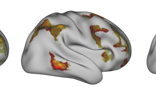
 Illustration of an individual performing a language task: predicted activity (yellow), recorded activity (red)UNIVERSITY OF OXFORD; IDO TAVOR, SAAD JBABDIEach of us has a unique neural connectome, a network within and between different brain regions that is preserved both at rest and during a range of activities. Now, scientists from the University of Oxford, U.K., and their colleagues have shown that this map of the brain’s activity at rest provides information on what that brain looks like when performing a task. The team’s results were published today (April 7) in Science.
“We show that we can essentially predict how people will use their brains based on [their resting brain activities],” said study coauthor Saad Jbabdi of Oxford.
Many neuroimaging studies pool data across individuals to identify brain activities on average. Jbabdi and his colleagues instead analyzed individual brain activity data from 98 Human Connectome Project participants. The brains of these individuals were previously scanned via functional magnetic resonance imaging (fMRI) while the participants were at rest and while they were performing 47 different tasks (including social interactions and memory-related tasks). Jbabdi’s team also analyzed images of the architecture of the individuals’ brains mapped using MRI and diffusion-weighted MRI, a technique that maps the flow of water molecules throughout the brain.
Individual brains differ in shape, architecture, ...


