
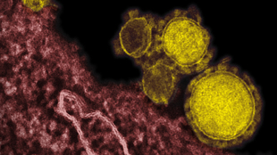
 Micrograph of MERS-CoV particles (yellow)FLICKR, NIAIDLast week, Saudi Arabian officials announced the 105th Middle East respiratory syndrome (MERS)-related death in the country since the causative coronavirus (CoV) emerged in the country in September 2012. While at first the MERS-CoV was largely a mystery, scientists have since been working to scan its genome, decipher its transmissibility, identify potential vectors, and develop vaccines against it. This week, two independent teams have advanced scientists’ understanding of the coronavirus: one group cultured the MERS-CoV, while another identified human antibodies that bind it and could inform therapies.
In February, Columbia University’s Ian Lipkin and his colleagues showed that MERS-CoV was common in camels living near areas where most of the documented human infections had occurred. Now, Lipkin’s team has identified diverse MERS-CoV quasispecies from Saudi Arabian camels. The researchers’ work was published in mBio this week (April 29).
“There appears to be a wide range of different viruses present within camels,” Lipkin told the University of Minnesota Center for Infectious Disease Research and Policy’s CIDRAP News.
“The finding of infectious virus strengthens the argument that dromedary camels are reservoirs for MERS-CoV,” coauthor Thomas Briese, also of Columbia University, said in a statement. “The narrow range of MERS viruses in humans and a very broad range in camels may explain in part why ...


