
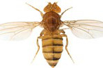
 UCSD, VALENTINO GANTZ AND ETHAN BIERResearchers from the University of California, San Diego, have developed an approach that utilizes CRISPR/Cas9 technology in Drosophila melanogaster to develop homozygous mutants in half the time it would take using traditional crosses. The team described its approach in Science this week (March 19).
“The study is well done and also very elegant,” said Ji-Long Liu of the University of Oxford who was not involved in the research. Liu called the method “a really clever way to . . . make the magic happen.”
 OZ OZKAYA, INSTITUTO GULBENKIAN DE CIENCIAA team led by investigators at Portugal’s Instituto Gulbenkian de Ciencia (IGC) has found that, following streptomycin treatment in mice, delivering a quorum-sensing molecule called pan-species autoinducer-2 (AI-2) to the animal’s guts can help reestablish beneficial bacterial populations there. The group’s results appeared in Cell Reports this week (March 19).
“While the vast majority of studies on the microbiome identify different bacteria present in the gastrointestinal tract, what is different and important about [this] approach . . . is in trying to manipulate the signaling in the GI tract,” said William Bentley, a bioengineer at the University of Maryland who was not involved in the work.
“It’s a proof of ...


