
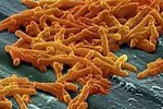
 WIKIMEDIA, CJC2NDClostridium difficile infections can be prevented through the rebalancing of bile acids in the gut by introducing certain commensal microbes, according to a study published in Nature this week (October 23). Eric Pamer of the Memorial Sloan Kettering Cancer Center in New York City and his colleagues have demonstrated the efficacy of this approach in both mice and humans.
“Lots of people have looked at using bacteria to mediate the so-called colonization resistance to C. difficile,” Vincent Young, a microbiologist and infectious disease physician at the University of Michigan who was not involved in the study, told The Scientist, “but this paper really goes a long way towards defining a good mechanism for how it happens.”
 HHMI, BETZIG LABNobel Laureate Eric Betzig and his colleagues described a new technique, lattice light-sheet microscopy, in Science this week (October 23). The approach hinges on illuminating thin sections of living sample one at a time using a targeted plane of light, enabling researchers to track the movements of single molecules in 3-D over time.
“With this microscope, I feel like Galileo,” Betzig told The Scientist. “No matter where we point it, we make a discovery, and we see something of incredible beauty.”
 HARVARD'S WYSS INSTITUTEBoston University’s James Collins and his colleagues have successfully freeze-dried gene networks and later rehydrated them, finding they were biologically active. Their work was published in Cell this week (October 23).


