
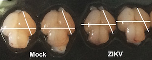
 Brains of fetal mice infected with Zika (ZIKV) and unifected mice (Mock)C. LI ET AL., CELL STEM CELLA trio of mouse studies published today (May 11) provides some of the strongest evidence yet that Zika virus infection can cause birth defects. Researchers at the University of São Paulo in Brazil and their colleagues reported in Nature that the Brazilian strain of the virus could cross the placentas of pregnant mice, resulting in microcephaly in developing embryos. Meanwhile, researchers from the Chinese Academy of Sciences reported similar findings in Cell Stem Cell after having studied a different infection route. And researchers at the Washington University School of Medicine in St. Louis found that Zika can infect pregnant, immune-deficient mice and their fetuses, causing restricted growth and fetal death; their findings were published in Cell.
“These papers are the link that was missing,” Shannan Rossi of the University of Texas Medical Branch (UTMB), who was not involved in the studies, told The Scientist. “Up until this point, there was no smoking gun [that Zika causes microcephaly]. Now we have a gun smoking.”
Zika was originally isolated from Rhesus monkeys in Uganda in 1947. A growing body of evidence suggests that the recent spike in cases of microcephaly in Brazil were caused by infection with an Asian strain of the virus primarily spread by Aedes aegypti mosquitoes. The virus has been shown to infect neural progenitor cells in vitro, and has also been found in the amniotic fluid of infected pregnant women and in fetal brains and retinas. But until now, ...


