
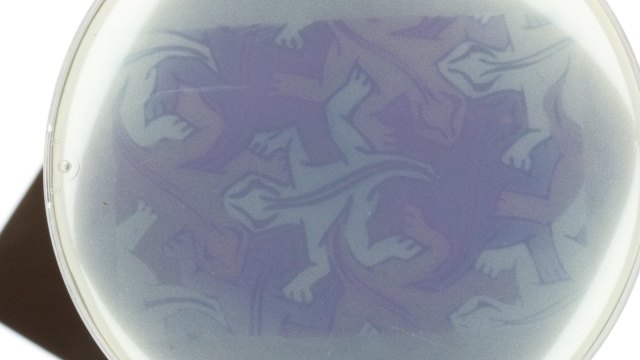
 FELIX MOSERMore than a decade ago, MIT researchers, led by biological engineer Christopher Voigt, engineered bacterial cultures to produce black-and-white photo negatives mirroring patterns of light projected onto the dish. Augmenting the cells to sense light and produce black pigment in response required only four genes. Now wielding a suite of 18 genes, Voigt’s team has created a microbial color palate that can paint a fruit still life, a geometric lizard motif, and a leaping Super Mario. The results appear in Nature Chemical Biology today (May 22).
“We think of it as sort of ‘disco bacteria,’” Voigt said. “You can imagine different light flashing depending on what the cells need to do.”
To engineer bacteria that react to certain wavelengths of light by producing a particular pigment, Voigt’s team leveraged several cutting-edge pieces of technology, including optogenetic laser light sensors, CRISPR-based gene-expression control, and the lab’s very own bacterial programming language. The result was a microscopic Rube Goldberg machine, which sprang into action when the researchers shined colored light onto one of their engineered E. coli cells.
 FELIX MOSERWavelength-specific light sensor molecules studding the surface of the cell switched on, activating dedicated genetic circuitry that ultimately spit out a desired product. In this “photography” experiment, the products were pigment molecules of the same color as the light input—red, green, or blue—which caused the bacterial photo paper to mimic the image projected upon it.
The utility of the system goes beyond making pretty pictures. ...


