“Breakthrough” Tough to Reproduce

An independent group could not replicate the results of a highly cited heart regeneration protocol, while others say they have succeeded.

Written byKerry Grens
| 4 min read

Register for free to listen to this article
Listen with Speechify
0:00
4:00
Share

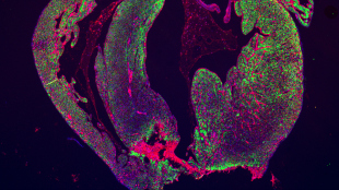
The resected area of a mouse heart is still missing and scar formation (red) is seen in the border of the resection line.STEM CELL REPORTS, ANDERSEN ET AL.In 2011, researchers reported having successfully induced the hearts of newborn mice to regenerate after a surgery. The feat was a major accomplishment in demonstrating the ability of a mammalian heart to regenerate, and the publication has been cited more than 300 times. But in a study published today (April 3) in Stem Cell Reports, a separate team tested the protocol and could not reproduce the findings.

“I haven’t seen one heart that has been regenerated in our lab,” said Ditte Andersen, the lead author of the new study and a researcher at the University of Southern Denmark. Andersen told The Scientist that she doesn’t have an explanation for the discrepancy. “We’ve discussed everything,” she said, but her team could not find a reason.

Hesham Sadek, the senior author of the 2011 paper and a researcher at the University of Texas Southwestern Medical Center in Dallas, read Andersen’s report and said the problem is technical. “I think that their techniques are flawed,” he said, although he would not provide further detail.

Sadek’s procedure involved cutting out about 15 percent of the heart at ...

Interested in reading more?

Become a Member of

The Scientist Logo
Receive full access to more than 35 years of archives, as well as TS Digest, digital editions of The Scientist, feature stories, and much more!
Already a member? Login Here

Related Topics

Meet the Author

  • kerry grens

    Kerry served as The Scientist’s news director until 2021. Before joining The Scientist in 2013, she was a stringer for Reuters Health, the senior health and science reporter at WHYY in Philadelphia, and the health and science reporter at New Hampshire Public Radio. Kerry got her start in journalism as a AAAS Mass Media fellow at KUNC in Colorado. She has a master’s in biological sciences from Stanford University and a biology degree from Loyola University Chicago.

    View Full Profile
Share
Illustration of a developing fetus surrounded by a clear fluid with a subtle yellow tinge, representing amniotic fluid.
January 2026, Issue 1

What Is the Amniotic Fluid Composed of?

The liquid world of fetal development provides a rich source of nutrition and protection tailored to meet the needs of the growing fetus.

View this Issue
Skip the Wait for Protein Stability Data with Aunty

Skip the Wait for Protein Stability Data with Aunty

Unchained Labs
Graphic of three DNA helices in various colors

An Automated DNA-to-Data Framework for Production-Scale Sequencing

illumina
Exploring Cellular Organization with Spatial Proteomics

Exploring Cellular Organization with Spatial Proteomics

Abstract illustration of spheres with multiple layers, representing endoderm, ectoderm, and mesoderm derived organoids

Organoid Origins and How to Grow Them

Thermo Fisher Logo

Products

Brandtech Logo

BRANDTECH Scientific Introduces the Transferpette® pro Micropipette: A New Twist on Comfort and Control

Biotium Logo

Biotium Launches GlycoLiner™ Cell Surface Glycoprotein Labeling Kits for Rapid and Selective Cell Surface Imaging

Colorful abstract spiral dot pattern on a black background

Thermo Scientific X and S Series General Purpose Centrifuges

Thermo Fisher Logo
Abstract background with red and blue laser lights

VANTAstar Flexible microplate reader with simplified workflows

BMG LABTECH