CRISPR Improves Disease in Adult Mice

Three groups of researchers used the gene-editing method to restore a protein deficient in Duchenne muscular dystrophy.

Written byKerry Grens
| 2 min read

Register for free to listen to this article
Listen with Speechify
0:00
2:00
Share

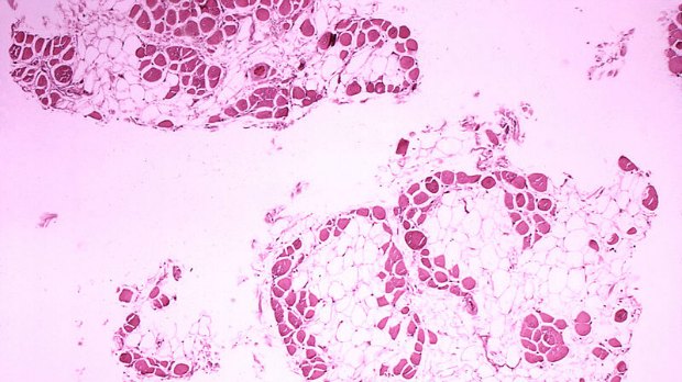
WIKIMEDIA, CENTERS FOR DISEASE CONTROL AND PREVENTION'S PUBLIC HEALTH IMAGE LIBRARYCRISPR has fixed the protein problems in adult mice that lie at the root of Duchenne muscular dystrophy (DMD), a progressive disease that saps kids of muscle strength and ultimately shortens their lives. Scientists had succeeded in using the gene-editing technique to restore protein function in human cells or mouse embryos, but this is the first time adult animals have been treated.

“The hope for gene editing is that if we do this right, we will only need to do one treatment,” Duke University’s Charlie Gersbach, who led one of three independent research teams that published results in Science last week (December 31), told The New York Times. “This method, if proven safe, could be applied to patients in the foreseeable future.”

The problematic protein is called dystrophin. All three groups took the same approach, first demonstrated in mouse embryos by Eric Olson of the University of Texas Southwestern Medical Center in 2014, to correct dystrophin deficiencies. They clipped a mutant exon from the gene for dystrophin, resulting in a truncated but functional protein. “Importantly, in principle, the same strategy can be applied to numerous types of ...

Interested in reading more?

Become a Member of

The Scientist Logo
Receive full access to more than 35 years of archives, as well as TS Digest, digital editions of The Scientist, feature stories, and much more!
Already a member? Login Here

Related Topics

Meet the Author

  • kerry grens

    Kerry served as The Scientist’s news director until 2021. Before joining The Scientist in 2013, she was a stringer for Reuters Health, the senior health and science reporter at WHYY in Philadelphia, and the health and science reporter at New Hampshire Public Radio. Kerry got her start in journalism as a AAAS Mass Media fellow at KUNC in Colorado. She has a master’s in biological sciences from Stanford University and a biology degree from Loyola University Chicago.

    View Full Profile
Share
Illustration of a developing fetus surrounded by a clear fluid with a subtle yellow tinge, representing amniotic fluid.
January 2026, Issue 1

What Is the Amniotic Fluid Composed of?

The liquid world of fetal development provides a rich source of nutrition and protection tailored to meet the needs of the growing fetus.

View this Issue
Skip the Wait for Protein Stability Data with Aunty

Skip the Wait for Protein Stability Data with Aunty

Unchained Labs
Graphic of three DNA helices in various colors

An Automated DNA-to-Data Framework for Production-Scale Sequencing

illumina
Exploring Cellular Organization with Spatial Proteomics

Exploring Cellular Organization with Spatial Proteomics

Abstract illustration of spheres with multiple layers, representing endoderm, ectoderm, and mesoderm derived organoids

Organoid Origins and How to Grow Them

Thermo Fisher Logo

Products

Brandtech Logo

BRANDTECH Scientific Introduces the Transferpette® pro Micropipette: A New Twist on Comfort and Control

Biotium Logo

Biotium Launches GlycoLiner™ Cell Surface Glycoprotein Labeling Kits for Rapid and Selective Cell Surface Imaging

Colorful abstract spiral dot pattern on a black background

Thermo Scientific X and S Series General Purpose Centrifuges

Thermo Fisher Logo
Abstract background with red and blue laser lights

VANTAstar Flexible microplate reader with simplified workflows

BMG LABTECH