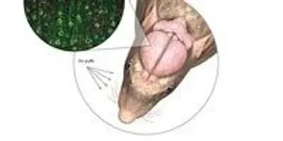
 CAUGHT IN THE ACT: Feng and colleagues recorded neuronal activity in mice engineered to express the calcium indicator GCaMP3 or GCaMP2.2 upon neuronal activation. Previously inserted cranial windows located over the motor cortex allowed the researchers to use fluorescent microscopy to examine the activation of specific neuron populations when the animals’ whiskers were stimulated by puffs of air. Green signals neuronal GCaMP3 expression; red marks neurons; and blue marks the nuclei of all brain cells.GEORGE RETSECK; NEURON IMAGE COURTESY OF QIAN CHEN AND GUOPING FENG
Observing the activity of specific neuronal circuits in the living brain can provide insight into how the brain functions normally and how it is affected by disease, psychological disorder, or injury. For that purpose, Guoping Feng, from the Massachusetts Institute of Technology, and colleagues have engineered transgenic mice whose neurons glow green when activated.
Neuronal activation leads to a rapid spike in intracellular calcium—a fact that prompted the development of fluorescent calcium dyes. But while these dyes provide an excellent readout of neuronal activity, says Feng, “you cannot put dyes into a specific population of neurons. . . . [And] you cannot image the same neurons over a long period of time.”
To get around these shortcomings, researchers have developed genetically encoded calcium indicators such ...


