Opinion: Brain Scans in the Courtroom

Advances in neuroimaging have improved our understanding of the brain, but the resulting data do little to help judges and juries determine criminal culpability.

Written byAndreas Kuersten
| 5 min read

Register for free to listen to this article
Listen with Speechify
0:00
5:00
Share

WIKIMEDIA; UPPSALA UNIVERSITY HOSPITAL, MIKAEL HÄGGSTRÖMA group of men exit a city hotel and begin making their way to a waiting car. As they approach the vehicle, another man opens fire on the group from 10 feet away, intending to kill one of them. Four men are wounded before the attacker is subdued by bystanders.

Imagine you’re a juror in the shooter’s trial for attempted murder.

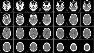
At trial, the shooter’s legal team pursues the insanity defense. The defense argues that, at the time of the offense, the accused—due to mental disease or defect—lacked substantial capacity to appreciate the wrongfulness of his actions or to conform to the requirements of the law. In support of this contention, the defense presents you and your fellow jurors with an image of the accused’s brain from a computerized axial tomography (CAT) scan. Accompanying this picture is the testimony of a defense expert, a neuroradiologist, saying that the shooter’s brain has relatively enlarged sulci and ventricles, signifying shrinkage and decay. A second defense expert, a psychiatrist, testifies that these characteristics are associated with schizophrenia.

To ...

Interested in reading more?

Become a Member of

The Scientist Logo
Receive full access to more than 35 years of archives, as well as TS Digest, digital editions of The Scientist, feature stories, and much more!
Already a member? Login Here

Related Topics

Meet the Author

Share
Illustration of a developing fetus surrounded by a clear fluid with a subtle yellow tinge, representing amniotic fluid.
January 2026, Issue 1

What Is the Amniotic Fluid Composed of?

The liquid world of fetal development provides a rich source of nutrition and protection tailored to meet the needs of the growing fetus.

View this Issue
Skip the Wait for Protein Stability Data with Aunty

Skip the Wait for Protein Stability Data with Aunty

Unchained Labs
Graphic of three DNA helices in various colors

An Automated DNA-to-Data Framework for Production-Scale Sequencing

illumina
Exploring Cellular Organization with Spatial Proteomics

Exploring Cellular Organization with Spatial Proteomics

Abstract illustration of spheres with multiple layers, representing endoderm, ectoderm, and mesoderm derived organoids

Organoid Origins and How to Grow Them

Thermo Fisher Logo

Products

Brandtech Logo

BRANDTECH Scientific Introduces the Transferpette® pro Micropipette: A New Twist on Comfort and Control

Biotium Logo

Biotium Launches GlycoLiner™ Cell Surface Glycoprotein Labeling Kits for Rapid and Selective Cell Surface Imaging

Colorful abstract spiral dot pattern on a black background

Thermo Scientific X and S Series General Purpose Centrifuges

Thermo Fisher Logo
Abstract background with red and blue laser lights

VANTAstar Flexible microplate reader with simplified workflows

BMG LABTECH