Single-Cell Suck-and-Spray

A nanoscopic needle and a mass spectrometer reveal the contents of individual cells.

Written byRuth Williams
| 3 min read

Register for free to listen to this article
Listen with Speechify
0:00
3:00
Share

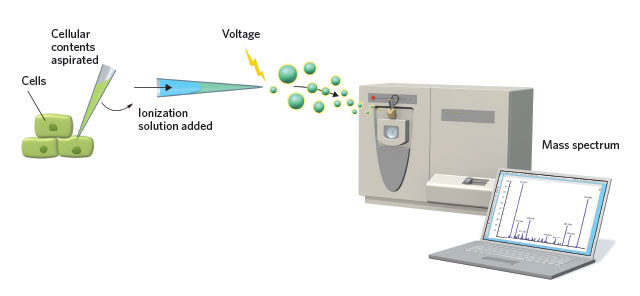
SINGLE-CELL MASS SPEC: To analyze the small-molecule components of an individual cell, a tiny capillary needle sucks material from the cytoplasm. After mixing the contents with an ionization solution, an applied voltage ejects the material from the needle and sends it into a mass spectrometer for analysis.
See full infographic: PDF
© GEORGE RETSECK

Analyzing the molecular constituents of a homogenized piece of animal or plant tissue may provide clues about the nature of the component cells’ functions. But it may also obscure striking functional differences between individual cells.

Not surprisingly, then, the study of single cells is a burgeoning area of research. While single-cell genomics and transcriptomics have benefited from DNA’s and RNA’s inherent ability to be amplified, single-cell proteomics and metabolomics must rely solely on miniscule quantities of starting material.

Researchers have thus been developing a variety of techniques for tackling such tiny volumes. The teams of Tsutomu Masujima at Hiroshima University and RIKEN in Osaka, Japan, for example, have been perfecting a mass spectrometry protocol for examining plant-cell metabolites that requires micromanipulation tools similar to those ...

Interested in reading more?

Become a Member of

The Scientist Logo
Receive full access to digital editions of The Scientist, as well as TS Digest, feature stories, more than 35 years of archives, and much more!
Already a member? Login Here

Related Topics

Meet the Author

  • ruth williams

    Ruth is a freelance journalist. Before freelancing, Ruth was a news editor for the Journal of Cell Biology in New York and an assistant editor for Nature Reviews Neuroscience in London. Prior to that, she was a bona fide pipette-wielding, test tube–shaking, lab coat–shirking research scientist. She has a PhD in genetics from King’s College London, and was a postdoc in stem cell biology at Imperial College London. Today she lives and writes in Connecticut.

    View Full Profile

Published In

Share
Illustration of a developing fetus surrounded by a clear fluid with a subtle yellow tinge, representing amniotic fluid.
January 2026, Issue 1

What Is the Amniotic Fluid Composed of?

The liquid world of fetal development provides a rich source of nutrition and protection tailored to meet the needs of the growing fetus.

View this Issue
Skip the Wait for Protein Stability Data with Aunty

Skip the Wait for Protein Stability Data with Aunty

Unchained Labs
Graphic of three DNA helices in various colors

An Automated DNA-to-Data Framework for Production-Scale Sequencing

illumina
Exploring Cellular Organization with Spatial Proteomics

Exploring Cellular Organization with Spatial Proteomics

Abstract illustration of spheres with multiple layers, representing endoderm, ectoderm, and mesoderm derived organoids

Organoid Origins and How to Grow Them

Thermo Fisher Logo

Products

Brandtech Logo

BRANDTECH Scientific Introduces the Transferpette® pro Micropipette: A New Twist on Comfort and Control

Biotium Logo

Biotium Launches GlycoLiner™ Cell Surface Glycoprotein Labeling Kits for Rapid and Selective Cell Surface Imaging

Colorful abstract spiral dot pattern on a black background

Thermo Scientific X and S Series General Purpose Centrifuges

Thermo Fisher Logo
Abstract background with red and blue laser lights

VANTAstar Flexible microplate reader with simplified workflows

BMG LABTECH