TK
| 2 min read
Researchers are using CRISPR for precise genetic manipulation of human-associated microbes as a promising avenue for improving human health.

infographic

Infographic showing the process of tail regeneration in tadpoles

Infographic: How Tadpoles Use Glucose to Fuel Tail Regrowth

Infographic showing transposable elements in cancer

Infographic: Transposable elements in cancer

Infographic comparing the fall and spring salmon runs

Infographic: An Incredible Journey

Illustration showing microscopic algae swim through mouse lungs and deliver nanoparticles of an antibiotic attached to their surfaces

Infographic: Algae Robots Transport Antibiotics to Infected Tissues

Timeline summarizing a series of petitions filed about the Chinook salmon

Timeline: An Extended Battle

Close-up shot of sea surface with small waves

The Constellation of Creatures Inhabiting the Ocean Surface

Neuston

Slideshow: Meet the Neuston, the Diverse Organisms Living at the Ocean’s Surface

Illustration showing where neuston reside

Infographic: Neuston Drift Atop the World’s Oceans

<em>Chlamydia</em> invades a host cell, forms a membrane-bound vacuole, or inclusion, and then modifies the protein composition of the structure&rsquo;s membrane. If immune cells detect <em>Chlamydia</em> before it forms the inclusion, they trigger T cells to produce interferon-&gamma; (IFN-&gamma;), a powerful cytokine. IFN-&gamma; activates the protein mysterin (also called RFN213), which attaches ubiquitin to the inclusion membrane, signaling the cell to destroy the inclusion&rsquo;s contents by dumping them into a lysosome (left). C. trachomatis produces GarD, a protein that integrates into the inclusion membrane itself and somehow prevents mysterin from attaching ubiquitin, allowing the bacterium to evade immune destruction while continuing to multiply and eventually bursting from the cell (right).

Infographic: How Chlamydia Evades Immune Detection

Infographic preview

Infographic: A Brain Implant Stops Tumor Growth in Rats

(Left) The optic lobes are thought to be involved in visual processing. In this study, researchers found that guppies with larger optic lobes more quickly learned a visual discrimination task&mdash;identifying which color well contained food. (Right) The fish telencephalon is thought to be involved in spatial learning, memory, and inhibitory control. Here, the researchers found that a larger telencephalon might enhance the fish&rsquo;s cognitive flexibility, allowing them to more quickly associate food with a new color after the researchers switched it.

Infographic: Two Guppy Brain Regions May Help Them Learn Tasks

Photo of a Guppy fish

Fish Brain Region Size Correlates with Cognitive Flexibility

3D imaging of organoid

Infographic: Generating Hundreds of 3D Organoid Images per Hour

Mind the Graph

Seeing Science: How to Visually Explain Complex Concepts

Genetic knockout experiments reveal a role for the protein in forming and maintaining synapses between motor neurons and muscle fibers in mice.

Infographic: Vangl2 in Muscles Keeps Neuromuscular Junctions Organized

Illustration showing assembly Versus Alignment

Infographic: The Sequencing and Assembly of the Human Genome

To flag neurons that have experienced genotoxic stress, researchers developed an in vivo sensor using an adeno-associated viral vector, called PRISM. Because a cell&rsquo;s DNA damage response (DDR)&mdash;which activates in response to stressors such as environmental toxins or the buildup of misfolded proteins&mdash;also responds to invading pathogens, PRISM has an easier time transfecting cells whose damage response mechanisms are preoccupied with existing DNA damage. Once inside, the virus hijacks the neuron&rsquo;s DNA replication machinery, which reverts an engineered frameshift mutation in the virus and thereby prompts the production of a fluorescent protein that can be observed via microscopy.

Infographic: DNA Damage Viewed with Unprecedented Clarity

Vaccine illustration&nbsp;

Infographic: Inducing Active Immunity Against Opioid Overdose

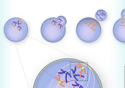
Infographic showing how bursting micronuclei promote cancer

Infographic: Chromosome Errors Cause Micronuclei and Drive Cancer

Illustration of a DNA-peptide conjugate molecule being pulled through a nanopore in a membrane.

Infographic: Reading Proteins with Nanopores

Trending

Photo of John Calhoun crouches within his rodent utopia-turned-dystopia

Universe 25 Experiment

Raised hand holding a gold medal against blue sky.

What Is Gene Doping in Sports, and How Can Experts Detect It?

An MRI image of a human brain, with a glowing red section which indicates a tumor.

Why Are Breast Cancers That Metastasize to the Brain Hard to Treat?

A female scientist looks at two white rodents in a clear plastic box.

Is This the End of Animal Testing? FDA Announces Plans to Phase Out Animals in Drug Safety Studies

Multimedia

Close up concept art of PCR wells with colorful contents

Improving Cell Therapy Sterility Testing

sartorius logo
Conceptual photograph of connecting the dots in a pathway through route planning.

The Computer-Assisted Retrosynthesis Revolution

Merck
May digest 2025 cover
May 2025, Issue 1

Study Confirms Safety of Genetically Modified T Cells

A long-term study of nearly 800 patients demonstrated a strong safety profile for T cells engineered with viral vectors.

View this Issue
iStock

TaqMan Probe & Assays: Unveil What's Possible Together

Thermo Fisher Logo
Meet Aunty and Tackle Protein Stability Questions in Research and Development

Meet Aunty and Tackle Protein Stability Questions in Research and Development

Unchained Labs
Detecting Residual Cell Line-Derived DNA with Droplet Digital PCR

Detecting Residual Cell Line-Derived DNA with Droplet Digital PCR

Bio-Rad
How technology makes PCR instruments easier to use.

Making Real-Time PCR More Straightforward

Thermo Fisher Logo

Products

fujirebio-square-logo

Fujirebio Receives Marketing Clearance for Lumipulse® G pTau 217/ β-Amyloid 1-42 Plasma Ratio In-Vitro Diagnostic Test

The Scientist Placeholder Image

Biotium Launches New Phalloidin Conjugates with Extended F-actin Staining Stability for Greater Imaging Flexibility

Leica Microsystems Logo

Latest AI software simplifies image analysis and speeds up insights for scientists

BioSkryb Genomics Logo

BioSkryb Genomics and Tecan introduce a single-cell multiomics workflow for sequencing-ready libraries in under ten hours