Toxin Wipes Out Persistent HIV

An antibody-toxin combo kills residual HIV hiding out in the organs of mice.

Written byKerry Grens
| 1 min read

Register for free to listen to this article
Listen with Speechify
0:00
1:00
Share

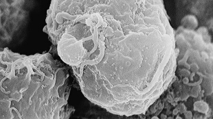
WIKIMEDIA, C. GOLDSMITHAlthough anti-retroviral therapy is quite good at tamping down the viral load in the blood of people infected with HIV, the virus can still hang out in tissues. So researchers designed an antibody that seeks out a viral envelope protein expressed by infected cells and tacked on to this infection-homing device a toxin that destroys the cell. They tested the therapy on HIV-infected model mice and published their results in PLOS Pathogens this week (January 9).

“Everywhere we look, the antibody is able to kill those infected cells,” University of North Carolina virologist J. Victor Garcia, a leader on the study, told the Los Angeles Times. In spleen, bone marrow, liver, lung, lymph nodes, and other areas, viral DNA dropped dramatically in mice administered the antibody-poison treatment, compared to animals given only conventional antiretroviral medications. “Our work provides evidence that HIV-infected cells can be tracked down and destroyed throughout the body,” Garcia said in a statement.

John Frater, an HIV researcher from the University of Oxford, told The Conversation: “The conclusions at this stage are that in this mouse model, [antiretroviral therapy] can be potentially improved with the addition of an immunotoxin. How this translates to human treatment is not known. ...

Interested in reading more?

Become a Member of

The Scientist Logo
Receive full access to more than 35 years of archives, as well as TS Digest, digital editions of The Scientist, feature stories, and much more!
Already a member? Login Here

Related Topics

Meet the Author

  • kerry grens

    Kerry served as The Scientist’s news director until 2021. Before joining The Scientist in 2013, she was a stringer for Reuters Health, the senior health and science reporter at WHYY in Philadelphia, and the health and science reporter at New Hampshire Public Radio. Kerry got her start in journalism as a AAAS Mass Media fellow at KUNC in Colorado. She has a master’s in biological sciences from Stanford University and a biology degree from Loyola University Chicago.

    View Full Profile
Share
Illustration of a developing fetus surrounded by a clear fluid with a subtle yellow tinge, representing amniotic fluid.
January 2026, Issue 1

What Is the Amniotic Fluid Composed of?

The liquid world of fetal development provides a rich source of nutrition and protection tailored to meet the needs of the growing fetus.

View this Issue
Skip the Wait for Protein Stability Data with Aunty

Skip the Wait for Protein Stability Data with Aunty

Unchained Labs
Graphic of three DNA helices in various colors

An Automated DNA-to-Data Framework for Production-Scale Sequencing

illumina
Exploring Cellular Organization with Spatial Proteomics

Exploring Cellular Organization with Spatial Proteomics

Abstract illustration of spheres with multiple layers, representing endoderm, ectoderm, and mesoderm derived organoids

Organoid Origins and How to Grow Them

Thermo Fisher Logo

Products

nuclera logo

Nuclera eProtein Discovery System installed at leading Universities in Taiwan

Brandtech Logo

BRANDTECH Scientific Introduces the Transferpette® pro Micropipette: A New Twist on Comfort and Control

Biotium Logo

Biotium Launches GlycoLiner™ Cell Surface Glycoprotein Labeling Kits for Rapid and Selective Cell Surface Imaging

Colorful abstract spiral dot pattern on a black background

Thermo Scientific X and S Series General Purpose Centrifuges

Thermo Fisher Logo