Most spectacularly evident in 2013 was how easily new techniques caught fire and spread to labs around the globe. Even researchers who don’t specialize in methods development are able to rapidly adopt—and improve upon—new approaches to answering their questions, and the result is an acceleration of progress and a cross pollination of disciplines. Here are some of the most exciting advances in the life sciences from 2013.
JINSONG LI
What can’t CRISPR do?
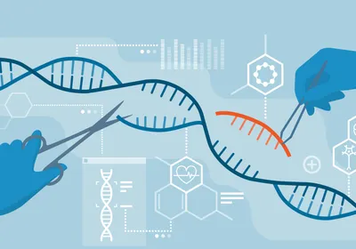
Clustered regularly interspaced short palindromic repeats, or CRISPR, is a tool used for genome editing. In tandem with an enzyme called Cas9, the CRISPR approach allows scientists to write the genetic code any which way they want. A few years ago, CRISPR was known only for its role in immunity in bacteria and archaea. Now, labs around the globe are wrapping their arms around the technique for myriad purposes.
“This new method is a game changer,”...
In 2013, advances in CRISPR applications decorated the pages of high profile journals with head-spinning frequency. In December alone, scientists demonstrated that CRISPR/Cas9 can correct disease causing genetic defects in mammals and human stem cells. And two papers in Science just last week laid out ways to use CRISPR in genomic screens.
MADELINE A. LANCASTEROrganoids galore
New types of lab-grown organ buds, also called organoids, popped up in 2013. In August, scientists raised small, three-dimensional models of embryonic human brains that could form some of the complex structures of the organ. “This demonstrates the enormous self-organizing power of human cells,” Jürgen Knoblich from the Institute of Molecular Biotechnology of the Austrian Academy of Science told The Scientist when his results were published.
In November, American and Spanish researchers published their data on functional, renal progenitor-like cells developed from human stem cells. A few months earlier, scientists reprogrammed human induced pluripotent stem cells (iPSCs) into liver buds that also took on three dimensions. The liver buds had seemingly normal metabolism and even hooked up to the host’s circulatory system when implanted in a mouse.
Grow-your-own organs may not be so far off. “If you could use iPSCs to generate a truly functioning organ, then you would have this unlimited suitcase of spare parts that would be genetically matched to individuals,” Stephen Duncan, the director of the Regenerative Medicine Center at the Medical College of Wisconsin, told The Scientist in July.
COURTESY RICARDO ROSSELLOPotent stem cells
The ability to reprogram skin cells to pluripotent stem cells by a simple gene-expression formula opened up a world of new experiments. This year, researchers continued to push ahead with better and faster methods to reinvent the identity of cells. Several months ago, scientists in Israel found a way to overcome one of the biggest limitations of inducing stem cells—the technique’s inefficiency. Typically, just about one out of every 10 cells prodded toward pluripotency actually do what researchers want it to, but Jacob Hanna from Israel’s Weizmann Institute of Science and his colleagues disabled a gene that represses pluripotency and voilà: near-perfect efficiency. “I never believed we’d get to 100 percent,” Hanna told The Scientist in September. “This shows that the process of reprogramming need not be random and inefficient.”
The induction of pluripotent stem cells reached new lows, as it were, in 2013. Researchers were able to generate in vivo more primitive forms of stem cells that had been accomplished before. Another group reprogrammed cells from animals lower on the tree of life than mammals, including birds, fish, and insects. And yet another team skirted the usual step of inserting genes into cells and instead used small molecules to coax cells into a pluripotent state.
J. CONNELL ET AL., UNIVERSITY OF TEXAS AT AUSTINExpanded uses for 3-D printing
3-D printers can make everything from test tube racks to centrifuges to tonight’s dinner. So why not print out your own custom mini microbial universe? Researchers have developed a gelatin mold that can house bacteria in separate compartments. “It’s basically Jell-O with things suspended in it,” chemist and bioengineer Jason Shear from the University of Texas at Austin told The Scientist in October.
The laser from a 3-D printer then builds a little entrapment around the bacteria. Although the bacteria are held in place, signals from the cells can move through the gel. Aleksandr Ovsianikov of the Vienna University of Technology in Austria, who was not involved in the project, pointed out that the technique allows researchers to build the mold any way they want. “This is a tool which potentially allows you to cross-link your gel, dress up your gel with biomolecules, or create channels in the same way,” said Ovsianikov. “This is a tool which is much more than 3-D printing.”
M. CHOIHydrogel implants
In another brilliant example of manipulating scaffolded cells, researchers developed a hydrogel implant embedded with a fiber optic cable that can direct the activity of cells with light. In this case, the light stimulated cells in the hydrogel to suppress high blood sugar levels in diabetic mice.
The hydrogel not only delivers light, it can detect it as well when the cells express fluorescent proteins. Seok Hyun Yun at Harvard University explained in October that his group didn’t actually invent anything new. “We put together somewhat disconnected, beautiful technologies to make [them] work in a single system . . . and then found a way to make it all work inside the body,” he told The Scientist. The approach could give a new angle for developing cell-based therapies.
The technique certainly needs some tweaking. HeLa cells are potentially tumorigenic and it’s not clear how the hydrogel would perform in different settings.
WIKIMEDIA, METOCMacGuyver-style microfabrication
Not all in scientific progress must involve fancier methods to solving problems. In fact, a little ingenuity and some Scotch tape goes a long way, according to Raquel Perez-Castillejos, a biomedical engineer at the New Jersey Institute of Technology in Newark. Scientists can use photolithographic technology to build little wells for culturing cells, but Perez-Castillejos and her colleagues found that cutting shapes into Scotch tape stuck to a glass slide works just fine.
The team cut small rectangles and other shapes into the tape. They then peeled away the tape, leaving the shapes of tape on the glass and made a cast of the slide with a silicon-based polymer. Once the casts were hard, they could be taken off the slide, turned upside down and plated with cells for microfluidics experiments. “Sometimes we are trying to provide very high precision to problems that don’t require it, and we make it complicated for no reason,” Perez-Castillejos said in the March issue of The Scientist.
WIKIMEDIA, INFERISThe next generation
Up-and-coming in vitro fertilization techniques gave parents in Philadelphia their next generation, a baby boy born in May. Researchers at the University of Oxford used next-generation sequencing to create a method to check embryos for chromosomal abnormalities, gene mutations, and mitochondrial genome mutations.
“Next-generation sequencing improves our ability to detect these abnormalities and helps us identify the embryos with the best chances of producing a viable pregnancy,” said Dagan Wells, a molecular geneticist at the NIHR Biomedical Research Centre at the University of Oxford, in a statement. “Potentially, this should lead to improved IVF success rates and a lower risk of miscarriage.”
The fertility doctor in Pennsylvania who used the technique to screen little Connor Levy when he was still a ball of cells said he expects the test to take off.
Thumbnail image credit: Wikimedia, National Institute of Health.